

 English
 French
 Spanish
 Russian
 Korean
 Japanese
What Is the Use of Marigold Flower Extract Lutein in Fish Feed?
China is a major fishing country in the world, accounting for about 70% of global aquaculture production, and there are many varieties, many of them colorful, such as yellow-colored yellow catfish (Pelteobagrus fulvidraco), greater yellow croaker (Pseudosciaena crocea), Chinese soft-shelled turtle (Pelodiscus sinensis), sweetfish (Plecoglossus altivelis), yellow striped amberjack (Seriola aureovittata), golden pomfret (Trachinotus ovatus), yellowfin bream (Sparus latus Houttuyn), etc., red-colored shrimp, crab, red sea bream (Pagrosomus major), rainbow trout (Oncorhynchus mykiss), golden threadfish (Nemipterus virgatus), Atlantic salmon, brocarded carp, etc. all have their own specific body colors in natural water environments, and they also display different protective and mating colors under different ecological and physiological conditions.
Body color is not only an important feature for classifying fish, but also a measure of their health status. At the same time, whether the body color is normal or not also directly affects the price of commercial fish. Often, the fish and shrimp in natural rivers have bright colors, delicious meat, and high prices. However, under intensive and artificial breeding conditions, the breeding cycle is shortened, and the main source of nutrients comes from compounded feed. In addition, the effective pigment sources in the feed are few and unstable, and aquatic animals can only obtain a small amount of natural aquatic organisms, so they cannot obtain sufficient natural pigment sources. This results in the body color of the fish becoming lighter, such as the big yellow croaker, golden pomfret, and Chinese soft-shelled turtle “turning white”; red sea bream “turning gray”, spotted catfish (Ietalurus Punetaus), yellowhead fish “turning white”, “variegated” or “black”; eel (Monopterus albus), loach (Oriental weatherfish) “red”; carp (Cyprinidae), crucian carp “black”, etc., all of which are abnormal body colors that affect the commercial value of the fish.
People's pursuit of the taste of “wild” fish has promoted research on the application of natural pigments in fish feed, but it has also led to the addition of some prohibited substances. The “yellow sodium powder dyed yellow croaker incident” detected in Lianyungang, Yangzhou, Nanchang, Wenzhou and other places in 2011 is a case of merchants using yellow sodium powder to dye faded yellow croakers to pass them off as wild yellow croakers in order to obtain high profits. Sodium yellow is an industrial pigment commonly used for dyeing wood and furniture. Due to its strong covering properties, it is not easy to remove once dyed, and it also contains industrial impurities and heavy metals. The state has expressly prohibited the use of such chemicals in the food industry. Frozen fish dyed with sodium yellow will cause malignant lesions in multiple internal organs of the human body and seriously damage health if consumed after being heated at high temperatures. Subsequently, it was revealed that yellow croaker fish in Qingdao and Jilin had been dyed with lemon yellow or sunset yellow. Sunset yellow and lemon yellow are synthetic pigments that are permitted food additives in China.
However, the Chinese Hygiene Standards for Food Additives only specify that they can be used in a limited range of foods such as fruit juices, prepared wines, sweets, pastries, prawn crackers, and lactic acid drinks.
The aluminum color tablets in sunset yellow and lemon yellow contain aluminum. If vendors add them in large quantities, long-term consumption of such fish can cause mental retardation, especially in sensitive groups such as pregnant women, infants and young children. In addition, there have been frequent incidents of non-pigment-added substances, toxic and harmful dye substances, or unauthorized dye substances being indiscriminately used in poultry eggs and vegetables. The “Sudan red duck egg incident” that shocked the country in 2006, the “lemon yellow dyed steamed buns” in March 2011, and the “dyed blood swan incident” in August 2011 once again brought the safety of pigment additives to the forefront. The safe use of feed additives is closely related to and inseparable from food quality and safety. Therefore, in order to solve the industry hotspot problem of discoloration plaguing aquatic animals and ensure the safety of aquatic product quality, we must consider the safety of the added pigments for both aquatic animals and human health.
1 Chemical properties of natural xanthophyll
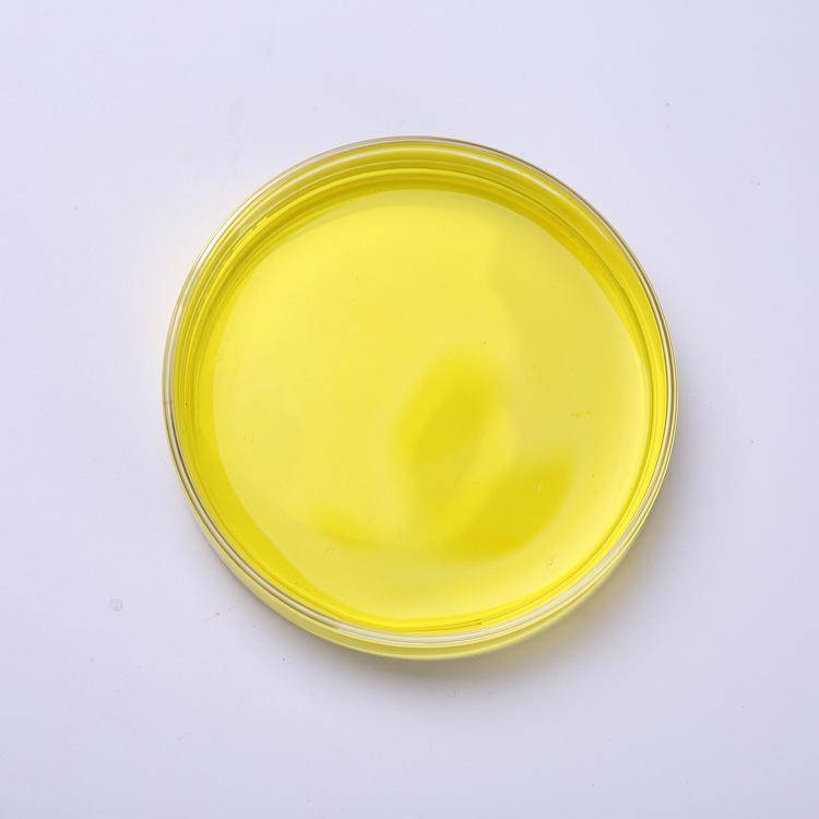
Natural xanthophyll is a class of non-vitamin A active dihydroxy oxygenated carotenoids widely found in vegetables, flowers, fruits, plants and egg yolks. which is most abundant in marigolds and is the main source of natural xanthophyll. It is orange-red or orange-yellow in color, and its main components are lutein and zeaxanthin. Of these, all-trans and cis-lutein and all-trans and cis-zeaxanthin account for 88%, and all-trans and cis-zeaxanthin accounts for 5%. It is insoluble in water, but soluble in oils and fatty solvents. Its molecular formula is C40H56O2, and its molecular structure is shown in Figure 1. Because it contains many conjugated double bonds in its structure, it is unstable to light, oxygen, and heat. However, the free hydroxyl group of lutein can be esterified with fatty acids to reduce its sensitivity to light and heat.
2. The scope of application of natural lutein as specified in Chinese food and feed
In food, the Ministry of Health of the People's Republic of China announced in 2007 that natural lutein extracted from marigold oil resin is approved as a new type of food additive. The properties are specified as orange to reddish orange, powdered, insoluble in water, and soluble in hexane. The scope and quantity of use are limited to 150 mg/kg in baked goods, 50 mg/kg in beverages (excluding packaged drinking water) (the amount in liquid drinks, and the amount in solid drinks is calculated according to the dilution multiple), 100 mg/kg in frozen foods, and 50 mg/kg in jellies and jams.
The content of each item must meet the following requirements: total carotenoids ≥ 80%, lutein ≥ 70%, zeaxanthin ≤ 9%. Subsequently, the Ministry of Health of the People's Republic of China announced in 2008 that Lutein Esters (lutein esters, Molecular Formula: C72H116O4, Molecular Weight: 1045.71) as a new resource food, with an edible amount of ≤12 mg/d. The scope of use includes baked goods, dairy products, beverages, ready-to-eat cereals, frozen drinks, condiments and sweets, but does not include infant foods. The lutein dipalmitate content is required to be >55.8%, and the zeaxanthin ester content <4.2%. Later, the scope of application of lutein as a food coloring agent derived from marigolds was expanded twice by the “Ministry of Health of the People's Republic of China Announcement” No. 1 of 2010 and the “Ministry of Health of the People's Republic of China Announcement” No. 16 of 2010, and the market demand continues to grow.

In feed, China's 2008 Catalogue of Feed Additive Varieties only allowed the use of natural lutein extracted from marigolds in poultry feed, and its use in aquatic animal feed had not yet been approved (Ministry of Agriculture of the People's Republic of China, Announcement 1126). In November 2010, the Ministry of Agriculture issued a solicitation for comments on the “Catalogue of Feed Additives (2010)” (draft for comments), which expanded the scope of use of natural marigold lutein to poultry and aquatic animals, while removing the use of chemically synthesized lutein in poultry.
3 Natural pigments and the body color of fish
Fish in nature have a variety of colorful body colors, and body color plays a vital role in the physiology, behavior and distribution of fish. The most common body color of fish is dark gray on the back and off-white on the belly. This is closely related to the living environment of the fish. The color of the back is similar to the color of the land, making it difficult for animals on land to see from top to bottom. The color of the belly is the same as the color of the water, making it difficult for enemies in the water to see from bottom to top. This is called protective coloration. Camouflage is especially important for fish living in the deep sea. For example, the rockfish relies on its body color being very similar to the color of the rocks to confuse the vision of other animals and protect itself or hunt prey. Like peacocks, some fish develop a brilliant marriage color during the breeding season to attract the opposite sex. For example, the male silver carp develops three bright orange-yellow stripes between the dorsal fin and the pectoral fin, accompanied by a pearl star at the operculum. The male blue mandarin fish has a distinct orange-red base on the pectoral fin.
In addition, body color is also a tool for aquatic animals to communicate and convey information. Squids and other animals use changes in body color to communicate within a group. The biological role played by the body color of fish is inseparable from various pigment cells and pigment particles. First, there are four main types of dendritic pigment cells in fish bodies: melanocytes, xanthocytes, erythrocytes, and iridocytes. Melanocytes are controlled by the nervous and endocrine systems, xanthophyl cells and red pigment cells are regulated by hormones, and iris cells are regulated by nerves. The first three pigment cells contain pigment particles, which absorb incident light of specific wavelengths to give the fish its various colors; the reflective layer of the iris cell can reflect light of a certain wavelength to give the fish its color. The main pigments in yellow chromatophores include lutein and zeaxanthin, canthaxanthin, cryptoxanthin, and tunaxanthin, which are carotenoids of the xanthophyll type, as well as zeaxanthin. Lutein is a type of carotenoid that is the main pigment in yellow chromatophores. Together with other pigments, it gives fish different colors and plays an important role in maintaining the characteristic colors of fish tissues such as muscles, fin rays, skin, gonads, and shells.
The types of carotenoids contained in the xanthophyl cells and/or red pigmented cells of different fish may differ. For example, Wang Anli et al. (2005) pointed out that red koi, black carp and scarlet carp can be separated into six pigment bands on thin-layer chromatography: light yellow, reddish orange, orange yellow, red, orange and apricot yellow; yellow writing koi has three pigment bands of light yellow, reddish orange and apricot yellow, while Showa tricolor koi has four pigment bands of light yellow, reddish orange, orange and apricot yellow. Thus, the main types of carotenoids in the skin, muscles or scales of aquatic animals can be colored with corresponding pigments to improve the body color of the animals. Each of the four pigment cells contains four pigment particles: melanin, carotenoids, pheophorbides and flavins. Under the action of neurogenic and/or hormonal regulation, each pigment particle undergoes physiological aggregation and diffusion and ecological numerical change and positional migration with myosin as the motor, so that the fish body displays different body colors. In addition to the number and distribution of pigment cells, the state of the pigment particles in the pigment cells, and the reflectivity of the reflective bodies in the iris cells, the body color of fish is also affected by the living environment, feed nutrition, the quality of the parents and young, age and gender, and physiological period.
4 Absorption and metabolism of natural lutein
Natural lutein contains lutein (lutein), which has a hydrophobic C40 isoprene carbon dioxide backbone, making it difficult to dissolve in chyme, but soluble in fats and fat-soluble solvents. Therefore, it needs to be assisted by the fats in food for digestion, absorption and metabolism. It is speculated that the absorption of lutein is similar to that of fat-soluble substances and in the small intestine. Sugawara et al. (2001) pointed out that the absorption of lutein, which is highly lipophilic, is four times that of fucoxanthin and neoxanthin, and that they can diffuse into the small intestinal villus epithelial cells with the help of lipid-soluble substances. In addition, the addition of a certain amount of lipid substances in food (or feed) can accelerate the intestinal absorption of lutein.
Sato et al. (2011) pointed out that the intestinal absorption of lutein can be significantly affected by the interaction of food components, and found that bile acids play a very important role in the intestinal absorption of lutein, which is known to be related to the absorption of fat-soluble substances (such as cholesterol) (NRC, 2011). According to domestic and foreign literature reports, the absorption process of marigold natural lutein is presumed to be as follows: in the chyme, lutein is emulsified into milk droplets along with the fat, and the milk droplets are further digested by lipase and bile. Lutein is finally solubilized in mixed colloidal particles, which are composed of bile acids, phospholipids, cholesterol, fatty acids and monoacylglycerol. The colloidal particles have a disc-like structure, surrounded by bile acids on the outside. These colloidal particles are then readily absorbed by the intestinal epithelial cells. Only a portion of the absorbed lutein is secreted into the lymphatic system in the form of chylomicrons and enters the blood circulation. The chylomicrons are then degraded by lipoprotein lipase, and the lutein in the chylomicron residue is absorbed by the liver.
The lutein absorbed by the liver is either stored in the liver or re-secreted into very low-density lipoproteins (VLDL) into the blood circulation, then delivered to low-density lipoproteins (LDL), and finally absorbed into tissues through LDL receptors. Lutein and zeaxanthin, which are highly lipophilic, are usually distributed in LDL and high-density lipoproteins (HDL) and are located on the outer surface of lipoprotein particles. Therefore, natural lutein is transported in both HDL and LDL. Thomas et al. also reported that zeaxanthin and lutein are mainly bound to HDL (53%), and are present in small amounts in LDL and VLDL (31% and 16%, respectively). Lutein absorbed into the blood is transported in the free form and stored in the form of lutein esters after transport to the tissues. Juliuszk et al. (1986) showed that lutein diesters transport lutein in the form of free alcohol in the blood of broilers. When the lutein content in the blood is too high, it is stored in the liver. Lutein in the serum samples of broilers was in the free form, while it was present in the form of a di-palmitate in the subcutaneous fat, indicating that only free lutein can enter the bloodstream of broilers and be stably stored after being converted to a di-palmitate in target organs.
There have been relatively few studies on the metabolism of non-VA-derived xanthophylls in mammals, and different researchers have detected different metabolites in the tissues of different animals. In mammals, the oxidation of the second hydroxy group to form a keto-carotene is the usual pathway for the metabolism of xanthophylls. Yonekura et al. found that the main metabolite of lutein in mouse tissue is keto-carotene. The main lutein metabolite in human plasma and retina is 3'-carboxy-lutein (i.e. 3-hydroxy-β,ε-carotene-3-1). Tyczkowski et al. found that 3'-carboxy-lutein, but not found in mouse tissue, which shows that the intestinal metabolites of lutein vary from species to species. Therefore, if the metabolism of natural lutein is to be clarified, researchers need to conduct studies on different species to obtain a final conclusion.
5 Application of natural lutein in fish
5.1 Coloring effect of natural lutein on fish
Animal coloring involves two stages: the saturation stage, which is achieved by deepening the yellow pigment, and the coloring stage, which enhances the color by adding red pigments to the yellow base. Each animal can only achieve the desired color by completing these two stages. Natural xanthophylls can color fish in two main ways: they can either be deposited directly in the scales, skin, adipose tissue and eggs, as in the case of the yellow catfish, bearded catfish, Chinese soft-shelled turtle, etc., or they can be converted into astaxanthin and then deposited in the tissues, as in Atlantic salmon, goldfish, red carp and koi carp.
Fish can synthesize melanin on their own, but they cannot synthesize carotenoids from scratch, so they must obtain carotenoids from different sources in their feed. Natural lutein extracted from marigolds is usually a bright yellow powder with strong coloring power. Its primary function is as a coloring agent, which can give aquatic animals and poultry yellow, yellowish brown, and golden yellow body colors. Leng Xiangjun et al. added natural lutein to the feed of about 52 g adult catfish and 3.5 g catfish fry to observe the coloring. They concluded that adding lutein products to the feed can effectively improve the body color of cultured catfish, and the appropriate additive amounts are 100 mg/kg feed (adult fish) or 50 mg/kg feed (fry). Wu Huachang et al. studied the effect of lutein on the body color of yellow catfish weighing about 52 g, and showed that when the natural lutein was added at a dosage of 100 mg/kg, it could effectively color the yellow catfish.
Shi Xiangyi et al. showed that adding 200 mg/kg lutein to the feed can effectively improve the body color of hybrid catfish within 20 days. Leng Xiangjun et al. added 150 mg/kg lutein to the goldfish feed, which effectively improved the goldfish body color. In addition, Olsen et al. (2006) found that lutein did not affect the deposition of astaxanthin when both were added to Atlantic salmon feed, and the skin appeared yellow. Li et al. showed that the order of skin coloration effects for the spotted fork-tailed worm was: lutein > zeaxanthin > astaxanthin > canthaxanthin > β-carotene. These studies show that although natural lutein derived from marigolds has a good coloring effect, Yasemen et al. used natural lutein to color the muscles of rainbow trout and found that the coloring effect was not as good as astaxanthin. The main reason may be that although rainbow trout has the ability to convert lutein into astaxanthin, the effect of directly using astaxanthin in terms of potency will be better. In addition, the main organ for lutein deposition is the skin, and lutein is only transferred to the muscles after the skin is saturated. However, the main organ for astaxanthin deposition is the muscle.
5.2 Other effects of natural lutein on fish
At present, many people believe that the addition of natural lutein to fish feed is only for coloring. However, as research continues, new functions of natural lutein from marigolds are being discovered. Research by Yang Wenping and others has shown that adding marigold extract to the feed helps improve the survival rate and growth rate of yellow catfish. An addition level of 0.8% can significantly increase the intestinal protease activity of yellow catfish, significantly increase the amylase activity of the stomach, intestines, liver and pancreas, and significantly increase the lipase activity of the intestines, liver and pancreas.
Yang Wenping et al. reported that the addition of 0.8% Jinbi Huang (marigold extract) to the feed can also significantly reduce the feed coefficient (P<0.05); the weight gain rate of the Jinbi Huang group is higher than that of the control group. Ding Xiaofeng et al. reported that the addition of the three pigments lycopene, canthaxanthin and marigold yellow to the feed had a certain effect on the fat content in the liver and pancreas of the fish. The fat content in the liver and pancreas of the fish in the canthaxanthin group decreased significantly by 18.2% compared to the control group.

Wang Lubo et al. showed that the addition of 24.2–1,700 mg/kg of natural lutein from marigolds to the feed significantly improved the growth performance of yellow catfish with an initial body weight of 21 g. A study on the wolf perch found that the added spirulina (which contains lutein) promoted growth by stimulating the activity of digestive enzymes in the pancreas and intestines, especially insulin. In a study using chlorella (containing lutein) to color six-gilled catfish, it was found that the group supplemented with chlorella was able to significantly increase the body length, weight and specific growth rate of the six-gilled catfish. In these studies, the application of marigold natural lutein to fish not only showed a coloring effect, but also promoted growth, increased the activity of digestive enzymes and to some extent reduced fat accumulation.
6 Safety of marigold natural lutein application
Safety evaluations of natural lutein have only been reported in rats, rhesus monkeys, and yellow croakers. Ravikrishna et al. found that in a 14-day acute toxicity test, rats given an oral dose of 2,000 mg/kg of Lutemax 2020TM (containing lutein and zeaxanthin) the rats did not show any toxic reactions or pathological abnormalities. Khachik et al. fed 2.8–4.4 kg rhesus monkeys 9.34 mg/kg lutein and 0.66 mg/kg zeaxanthin for 12 months, and it did not cause visual impairment.
Hariku et al. studied the toxicological model of free lutein and lutein esters extracted from marigold petals in Wistar male and female rats, and no negative effects were observed in either the short-term or long-term model. Wang Lubo et al. found that natural lutein extracted from marigolds is safe for reasonable use in aquatic animals. This generally shows that natural lutein derived from marigolds is safe as a feed additive and food additive. However, further research is needed on the use of natural lutein as an additive.


