What Is the Physiological Character of Phycocyanin?
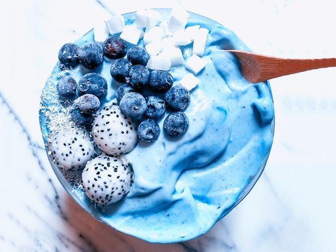
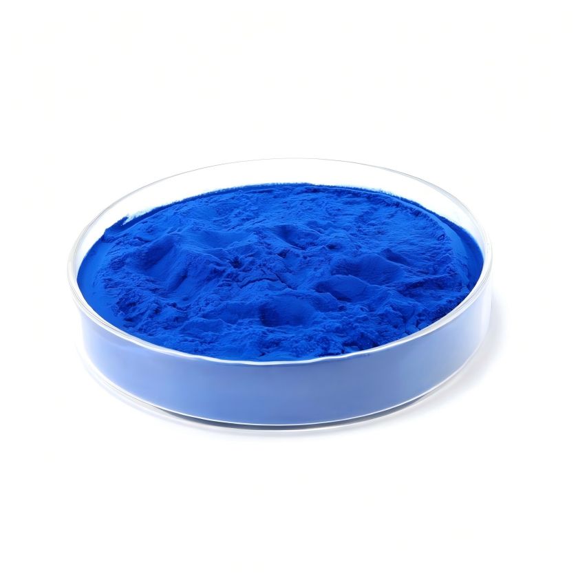
Phycocyanin (PC) is a photosynthetic pigment found in the cells of cyanobacteria and red algae that can efficiently capture light energy. It has a molecular weight of about 40 ku and consists of two subunits, α and β. One open-chain tetrapyrrole ring is covalently bound to the peptide chain. The isolated and purified water-soluble phycocyanin is blue in solution and emits purple fluorescence. It has a specific absorption peak at a wavelength of 620 nm, and its purity can be expressed as A620/A280. Due to its advantages of water solubility, non-toxicity, clarity and strong coloring power, phycocyanin is widely used as a food coloring agent and cosmetic additive. Phycocyanin has fluorescence and can be used as a fluorescent marker. In addition, studies have shown that phycocyanin has certain medical value. In recent years, it has been found that phycocyanin has a variety of biological activities, such as anti-cancer, anti-oxidation, treatment of cerebral ischemia damage, and improvement of immunity. Research on phycocyanin has become a research hotspot in natural marine drugs, and the author has conducted a review of its biological activities.
1 Research on the anti-tumor activity of phycocyanin
1.1 In vitro anti-tumor test of phycocyanin
It has been reported in literature both domestically and abroad that spirulina protein has a direct killing effect on tumor cells. Schwartz et al. [1] found that spirulina protein has an inhibitory effect on cancer cells. Wang Yong et al. [2] found that spirulina protein has a strong inhibitory effect on the growth of Hela cells cultured in vitro, and that when the concentration of spirulina protein in the culture system is increased from 10 mg/L to 80 mg/L, the inhibition rate gradually increases from 3.5% to 31.0%. Chen Xinmei et al. [3] found that when the appropriate dose of phycocyanin extract is used, it can directly inhibit the growth of cancer cell lines. It was also found that the maximum effective concentration of phycocyanin extract for inhibiting the growth of different cancer cells is different. The effective concentration for the liver cancer cell SMMC-7721 is 100 mg/L, which is much higher than the 50 mg/L for the laryngeal cancer cell HEp-2. indicating that the growth inhibition of laryngeal cancer cells is more sensitive to the influence of phycocyanin extract, and that the inhibitory effect at the same concentration is also different for different cells, which provides a basis for the development of phycocyanin extract as an anti-cancer agent.
Chen Tianfeng et al. [4] used ion exchange and gel filtration chromatography to purify selenium-containing phycocyanin from selenium-rich spirulina. The purified selenium-containing phycocyanin and phycocyanin have the effect of inhibiting the growth and proliferation of human malignant melanoma cells (A-375). The IC50 of the purified selenium-containing phycobiliprotein and phycobiliprotein is 44.5 and 65.9 μmol/L, respectively, which can significantly inhibit the proliferation of A-375 cells. In addition, phycobiliprotein has different degrees of inhibitory effects on human leukemia cell lines HL-60, K-562 and U-937 cells cultured in vitro, and there is a concentration-dose effect, with strong inhibitory effects at high concentrations. Due to the large molecular weight of phycocyanin, it is difficult for it to directly interact with cells. Zhang Xin et al. [5] obtained the active C-PC subunit through CM-Sepharose F.F. cation exchange chromatography, dialysis, and renaturation. The effects of C-PC, a and β subunits on the growth of the lung adenocarcinoma cell line SPC-A-1 were studied using a CCK-8 kit. The results showed that phycocyanin and its subunits have an inhibitory effect on the growth of this cell line, with the β subunit having the best effect, indicating that they have a certain antitumor potential.
1.2 Research on the physiological mechanism of the antitumor activity of phycocyanin
Li Bing et al. [6-7] believe that the mechanism of the antitumor effect of phycocyanin may be that phycocyanin, as a mitogen, binds to the mitogen receptor on the surface of tumor cells, which promotes the transcriptional expression of the CD59 gene in the cell through receptor cross-linking and protein kinase activation, inducing the activation of the death domain, thereby inhibiting the proliferation of tumor cells and promoting apoptosis of tumor cells.
In addition, in order to study the effect of phycocyanin on apoptosis of HeLa cells in vitro and its underlying mechanism, Li Bing et al. [8] first found by MTT assay that phycocyanin can inhibit the proliferation of HeLa cells and there is a concentration-dose effect. In order to gain a deeper understanding of whether this inhibitory effect is related to apoptosis induction, further research was carried out on the characteristics of the cells. Electron microscopy revealed that HeLa cells treated with phycocyanin showed obvious ultrastructural changes. The agarose gel electrophoresis structure showed that phycocyanin could induce DNA fragmentation in HeLa cells and produce ladder-like electrophoretic bands, which is a typical feature of cell apoptosis.
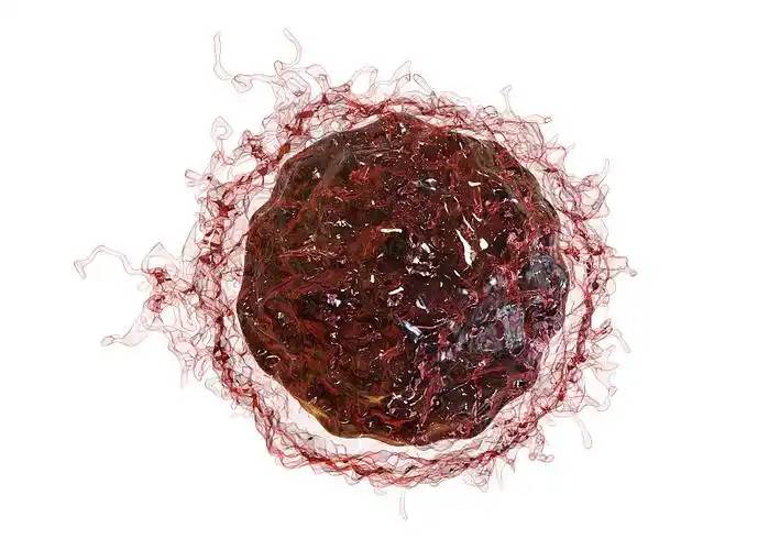
Subsequently, flow cytometry was used to detect the effect of phycocyanin on cell cycle changes and apoptosis in HeLa cells, and immunohistochemistry was used to further detect the effect of phycocyanin on the expression of apoptosis-related genes in HeLa cells. The results showed that phycocyanin can promote the expression of pro-apoptotic genes Fas and ICAM-1 and inhibit the expression of anti-apoptotic gene Bc1-2. Since caspase plays an important role in the apoptotic signaling pathway [9], the activity of caspase in HeLa cells treated with phycocyanin was further detected. The results showed that caspase-2, -3, -4, -6, -8, -9, and -10 were all activated, indicating that the apoptosis of HeLa cells induced by phycocyanin is caspase-dependent. Cytochrome C is an important respiratory chain protein that is released from mitochondria into the cytoplasm, which is a characteristic of cells undergoing apoptosis [10] and can be used as a criterion for determining the early stages of apoptosis. The results of the experiment showed that phycocyanin can induce the release of cytochrome C, which also confirms that phycocyanin achieves its goal of killing tumor cells by inducing apoptosis in HeLa cells.
COX-2 selective inhibitors can significantly inhibit tumor growth and tumor angiogenesis, induce apoptosis of tumor cells, and also enhance the cytotoxicity of chemotherapeutic drugs and the radiosensitivity of tumors. Reddy et al. [11] found that phycocyanin can selectively inhibit COX-2 activity to induce apoptosis of tumor cells. RAW264.7 phagocytes were stimulated with lipopolysaccharide (LPS). The results showed that phycocyanin had a dose-dependent inhibitory effect on the proliferation of this cell line.
2. Research on the antioxidant activity of phycocyanin
Domestic and foreign literature shows that natural phycocyanin and other phycocyanins both have antioxidant activity that scavenges hydroxyl radicals and hydrogen peroxide radicals [12-14]. Studies on the effect of phycobiliproteins on cytokines have found that phycobiliproteins can eliminate oxidative stress, thereby leading to TNF-a responses [15].
Chen Tianfeng et al. [4] used ion exchange and gel filtration chromatography to purify selenium-containing phycobiliproteins from selenium-rich spirulina. The antioxidant activity of selenium-containing phycobiliproteins was evaluated by comparing the scavenging abilities of selenium-containing phycobiliproteins and phycobiliproteins against four different free radicals, namely, the total antioxidant capacity, the ability to scavenge DPPH radicals, anionic peroxides and hydrogen peroxide-induced hemolysis, thus evaluating the antioxidant activity of selenium-containing phycobiliproteins. The results showed that the purified selenium-containing phycobiliprotein exhibited better antioxidant effects than phycocyanin at a specific dose in terms of its ability to scavenge ABTS, DPPH, anionic peroxides and AAPH radicals. After enrichment with organic selenium, phycobiliproteins can significantly improve their antioxidant properties, so the antioxidant research of selenium-enriched phycobiliproteins has received widespread attention in recent years [16-18].
Chenghua et al. [19] studied the selenium-enrichment ability of C-phycocyanin from marine Spirulina platensis and the scavenging effect of selenium-containing C-phycocyanin on superoxide anions and hydroxyl radicals. The results showed that when cultivated with the addition of a low concentration of selenium (40 to 80 mg/L), the selenium enrichment effect of marine Spirulina platensis C-phycocyanin was significantly stronger than that of freshwater Spirulina platensis C-phycocyanin. At a selenium concentration of 40 mg/L, C-phycocyanin had the highest selenium utilization rate and the largest selenium enrichment factor (0.9%); at a selenium concentration of 60 mg/L, C-phycocyanin had the highest selenium content (402 mg/kg). Selenium-containing C-phycocyanin has a stronger scavenging effect on superoxide anions and hydroxyl radicals than C-phycocyanin. The scavenging effect is positively correlated with the selenium content and protein concentration of C-phycocyanin. The selenium-containing C-phycocyanin group with the highest selenium content (402 mg/kg) has a scavenging rate of 83% and 35% for superoxide anions and hydroxyl radicals, respectively, at a concentration of 180 μg/ml. the removal rates of superoxide anions and hydroxyl radicals reached 83% and 35%, respectively, which is much higher than the removal effects of corresponding proteins from other freshwater species under the same conditions. The results of the study show that spirulina cultivated with seawater can significantly improve the selenium-enrichment capacity of C-phycocyanin and the activity of selenium-containing C-phycocyanin in removing superoxide anions.
Wang Xingping et al. [20] studied the antioxidant capacity of Spirulina platensis protein through non-lipid system antioxidant tests and in vitro antioxidant tests. The results showed that Spirulina platensis protein has a good quantitative-qualitative relationship with respect to scavenging oxygen free radicals, hydroxyl radicals and H2 O2. In an in vitro system, gexianmi phycocyanin significantly reduced the production of MDA and the levels of peroxides in the blood and liver, protecting cell membranes and red blood cells. It was shown that gexianmi phycocyanin has a protective effect on liver mitochondrial damage in mice and significantly affects the plasma's ability to scavenge reactive oxygen species.
3 Study of phycocyanin protecting islet cells
Zhang Rui et al. [21] established a type 2 diabetic rat model and used phycocyanin and metformin to treat the rats by gavage. The expression of nuclear factor κB (NFκB) and NFκB inhibitor protein (IκB) in islet cells was detected using immunohistochemistry. The results showed that after successful modelling in rats, fasting blood glucose increased significantly. After intervention and treatment, the fasting blood glucose of rats in the algin blue protein group and metformin group was significantly lower than that of the model group. The expression of NFκB in pancreatic islet cells of diabetic rats was significantly increased, while the expression of IκB was significantly decreased. After treatment with phycocyanin and metformin, the expression of NFκB in pancreatic islet cells of rats was significantly lower than that of the diabetic model group, while the expression of IκB was significantly higher than that of the diabetic group. This indicates that phycocyanin can inhibit the activity of the IκB/NFκB pathway in pancreatic islet cells of type 2 diabetic rats, and has a certain protective effect on pancreatic islet cells.
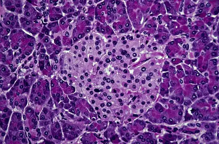
Ma Xuan et al. [22] also established a type 2 diabetes rat model. After the rats were successfully modeled, their body weight was significantly lower than that of the normal group, fasting blood glucose was significantly higher, the islets of Langerhans were atrophied, the structure was indistinct, the cells were disordered, the cytoplasm was swollen, there was vacuolar degeneration, etc., and the expression of islet cell iNOS was enhanced. After treatment with phycocyanin, fasting blood glucose was significantly lower than that of the model group, and the difference was significant. After treatment with phycocyanin and metformin, the expression of iNOS in pancreatic islet cells was significantly lower than in the diabetic model group, and the morphological structure of pancreatic islet cells was improved. This indicates that phycocyanin can inhibit the expression of iNOS in pancreatic islet cells of diabetic rats and has a protective effect on pancreatic islet cells.
4 Study of the anti-inflammatory activity of phycocyanin
Romay et al. [23] showed that phycocyanin exhibited anti-inflammatory effects in 12 inflammatory test models. Phycocyanin can scavenge alkyl, hydroxyl and peroxyl radicals, inhibit lipid peroxidation in microsomes induced by ferrous ions (Fe2+) and ascorbic acid, and its antioxidant effect is related to the inhibition of tumor necrosis factor (TNF) and NO production. Gonzalez et al. [24] used an enema of 1 ml 4% acetic acid to establish a colitis model, 150, 200, and 300 μg/kg of algin blue protein oral solution was given 30 minutes before, and MPO activity was measured 24 hours later. The colon tissue was examined histopathologically and by ultrastructure. The results showed that algin blue protein can reduce MPO activity. Microscopic observation showed that algin blue protein inhibits the invasion of inflammatory cells and reduces colon tissue damage. Vadiraja et al. [25] reported that Phycocyanin has a protective effect on CCl4-induced liver damage in mice, can almost restore the serum liver enzyme levels to those of the control group, has a protective effect on liver enzymes, and reduces the loss of 6-phosphogluconase and microsomal cytochrome P140.
5 Research on the hepatoprotective activity of phycocyanin
Suo Yourui et al. [26] established a rat model of alcoholic liver injury and found that administration of phycocyanin complex can effectively inhibit alcohol-induced liver cell damage. Compared with the model group, the serum ALT and AST levels of rats in the phycocyanin complex groups decreased, with significant decreases in the low-dose and high-dose groups. There was a significant dose-effect relationship, indicating that phycocyanin complex has a good effect of relieving hangover and protecting the liver. When rats were given the algin protein complex, all dose groups could significantly inhibit the lipid peroxidation reaction caused by alcohol, inhibit the increase in serum and liver MDA, protect liver cells, and the dose-effect relationship was significant. The algin protein complex can prevent the depletion of GSH caused by long-term alcohol consumption, and can significantly increase the activity of GSH, thereby protecting liver cells. Therefore, it can be seen that phycocyanin complex has the effect of detoxifying alcohol, inhibiting alcohol damage to liver cells, and protecting liver function. In addition, when rats are given alcohol and phycocyanin complex at the same time, it can effectively prevent and treat abnormal changes in tissues such as hepatic steatosis and punctate necrosis.
6 Immune regulation
Tang Mei et al. [27] confirmed through experiments that that phycocyanin can promote the effect of phytohaemagglutinin (PHA) in stimulating lymphocyte transformation, restore the ability of T cells to form E rosettes after being damaged by cyclophosphamide, and in particular, it has a good restorative effect on the formation of active E rosettes; it can also significantly increase the number of antibody-forming cells and the ability to produce antibodies in spleen cells from normal mice and mice with low immune function treated with hydrocortisone.
7 Conclusion
In summary, phycocyanin is a naturally occurring substance that is non-toxic and highly effective. It has a wide range of pharmacological activities, and its production, development and marketing provide favorable conditions for improving the income of local farmers and developing the economy. Exploring the sources of selenium, strengthening the development of selenium-rich agricultural products, and protecting selenium-rich soil are of great significance for maintaining the sustainable development of the local economy. Therefore, it is recommended to conduct a survey of the selenium content of local coal combustion, industrial emissions, irrigation water, etc., to determine the formation mechanism of local soil selenium enrichment.
References
[1] Chen Jiayou. On the vigorous cultivation of selenium-rich agricultural products [J]. China Science and Technology Information, 2005(7) : 33.
[2] Hao Libo, Qi Changmou. Geochemical Principles [M]. Beijing: Geological Publishing House, 2004.
[3] Hou Chuntang, Li Ruimin, Feng Cui'e, et al. Content and methods of regional agro-ecological geological survey—A case study of the Hebei Plain [J]. Geological Science and Technology Information, 2002, 21 (1): 66-70.
[4] Li Ruimin, Hou Chuntang. Progress in agricultural geology research at home and abroad [C] / / Jiang Jianjun, Hou Chuntang. New progress in agricultural geology research in China. Beijing: China Geodetic Publishing House, 2003.
[5] Li Yinan, Xue Liwen. Discussion on the standard of selenium content in selenium-rich health foods [J]. Guangzhou Trace Element Science, 2000, 7 (5): 4-6.
[6] Sun Wenguang, Liu Fengwu, Leng Xuyong, et al. Geological Survey Report on the Agricultural Ecological Environment of Tailai Basin, Shandong Province [R]. Jinan: Shandong First Institute of Geological and Mineral Exploration, 2009.
[7] Wang Jinghuai, Shi Chenzi. Discussion on the Development of Selenium-rich Agricultural Products and the Standard for Selenium Content [J]. Tianjin Agricultural and Forestry Science and Technology, 2005(3): 15-17.
[8] Wu Wanzheng, Wu Zhong. Trace element selenium and human health [J]. Guangdong Trace Element Science, 2000, 7 (11): 7-11.
[9] Yan Baowen, Li Jing. A preliminary discussion on agricultural environmental geology and agricultural environmental geological problems [J]. Hydrogeology and Engineering Geology, 2000(5): 41-43.
[10] Yan Peng, Xu Shiliang, Qu Kejian, et al. Shandong soil [M]. Beijing: China Agricultural Press, 1994.
[11] Zhang Zengqi, Liu Mingwei, Song Zhiyong, et al. Shandong rock strata [M]. Wuhan: China University of Geosciences Press, 1996.
[12] China National Environmental Monitoring Center. Background values of soil elements in China [M]. Beijing: China Environmental Science Press, 1999.
[13] Zou Jianjun, Li Shuanglin, Cheng Xinmin. Application of environmental geochemistry in agricultural environmental pollution [J]. Marine Geology Dynamics, 2004, 20(1): 30-33.


 English
 French
 Spanish
 Russian
 Korean
 Japanese