English
 French
 Spanish
 Russian
 Korean
 Japanese
Eliminating Heavy Metal Risks: Green Spring Technology's Phycocyanin for Worry-Free Formulations
In today's era where natural pigments and health ingredients are highly valued, phycocyanin has emerged as a popular choice in the food, health supplement, and cosmetics industries due to its unique blue hue and rich nutritional value. However, as market demand rapidly grows, raw material safety concerns have become increasingly prominent, with heavy metal contamination emerging as an “invisible killer” plaguing the entire industry.
Recent market monitoring data indicates that some phycocyanin sources of unknown origin carry risks of exceeding permissible levels of heavy metals like lead, cadmium, and mercury. These heavy metal contaminants not only compromise the pigment's color and stability but also pose potential health threats to consumers through product accumulation. A R&D director at a prominent beverage company confessed: “Heavy metals are our greatest concern in raw material procurement. A single instance of non-compliance could jeopardize an entire product line.” "
The root causes of this issue include:
· Water pollution in algal growth environments
· Non-standardized production and processing techniques
· Lack of stringent end-to-end quality control
“Heavy metal concerns not only compromise product safety but also hinder the healthy development of the entire industry,” stated the Quality Director of Green Spring Technology. “Many brands have had to abandon or postpone new product development plans due to the inability to secure stable, reliable low-heavy-metal raw materials.”
Fortunately, a breakthrough solution now addresses this industry challenge. Leveraging proprietary clean cultivation technology and purification processes, Green Spring Technology has successfully developed naturally compliant phycocyanin raw materials with heavy metal levels far below international standards, offering reliable options for companies prioritizing safety and quality.
We understand that raw material safety is the cornerstone of product innovation. Green Spring Technology is committed to enabling every client to eliminate heavy metal concerns through rigorous quality control and transparent testing systems, allowing them to focus on product innovation and market expansion.

Part One: Green Spring Technology Safeguards Safety and Quality
Green Spring Technology deeply understands the critical importance of heavy metal control for phycocyanin raw material quality. By establishing a comprehensive quality assurance system, we provide clients with truly safe and reliable product solutions.
1. Source Control: Prevention First
We firmly believe that superior products begin with pristine origins. Green Spring Technology has established independently controlled algae cultivation bases:
· Strict Environmental Selection: Sites are located in natural waters free from industrial pollution, with regular water quality monitoring.
· Ecological Cultivation: Closed photobioreactors eliminate external environmental contamination.
· Purified Strains: Multi-generation selection yields premium algae strains, ensuring quality at the genetic level.
“Our cultivation water meets drinking water standards—the first line of defense for raw material safety.” —Green Spring Technology Cultivation Base Manager
2. Innovative Processes for Precise Heavy Metal Removal
Green Spring Technology's patented purification technology achieves effective heavy metal control:
Chromatography Purification System
· Utilizes multi-stage column chromatography for selective adsorption of heavy metal ions
· Completes separation at low temperatures to preserve phycocyanin bioactivity
· Removal efficiency reaches 99.7%, far exceeding industry benchmarks
Membrane Separation Technology
· Utilizes ultrafiltration membranes with precise molecular weight cut-off
· Effectively separates heavy metal complexes
· Simultaneously removes other impurities
3. Authoritative Testing, Data-Driven Verification
We have established a quality inspection system exceeding national standards:
Testing Items and Standard Comparisons
Test Item | International Standard Limit | Green Spring Technology Actual Measurement Data |
Lead (Pb) | ≤1.0 mg/kg | ≤0.1 mg/kg |
Cadmium (Cd) | ≤0.5 mg/kg | ≤0.05 mg/kg |
Mercury (Hg) | ≤0.1 mg/kg | ≤0.01 mg/kg |
Arsenic (As) | ≤1.0 mg/kg | ≤0.2 mg/kg |
Certification Qualifications
· ISO 22000 Food Safety Management System Certified
· FDA GRAS (Generally Recognized as Safe) Certified
· EU Novel Food Certified
· Third-party testing reports provided for each batch
4. Dual Assurance: Prioritizing Naturalness and Safety
While strictly controlling heavy metal content, we equally emphasize preserving the product's natural properties:
Active Retention
· Protein content ≥85%
· Color value (A620/A280) ≥4.0
· Antioxidant activity retention ≥95%
Quality Stability
· Consistent performance throughout 36-month shelf life
· Batch-to-batch variation controlled within ±2%
· Suitable for diverse processing conditions
A partner client remarked: “After using Green Spring Technology's phycocyanin raw material, our products not only passed the most stringent export inspections but also received market acclaim for their outstanding color performance. This peace of mind is hard to find with other suppliers.”
Through end-to-end quality control and continuous technological innovation, Green Spring Technology enables clients to achieve true “ready-to-use” solutions, eliminating concerns about heavy metal issues.
Part Two: Application Scenarios
Green Spring Technology's low-heavy-metal phycocyanin raw material provides a safe and reliable foundation for innovation across industries, empowering the development of more competitive end products.
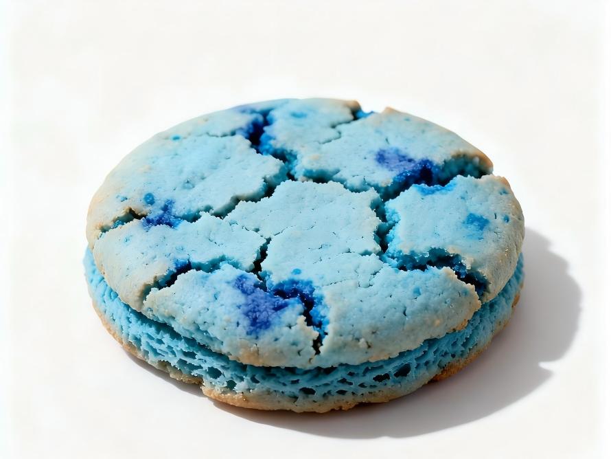
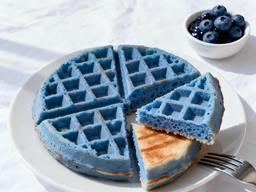
1. Food & Beverage: Redefining Natural Blue
In the food and beverage sector, safety is paramount in product development. Our raw material unlocks new possibilities for clients:
Innovative Case: Infant Formula
· An international brand incorporated our raw material into its new “Deep-Sea Algae Nutrient Rice Cereal”
· Successfully passed the most stringent heavy metal testing standards for infant foods
· Natural blue hue significantly enhanced product appeal, exceeding first-month sales projections by 200%
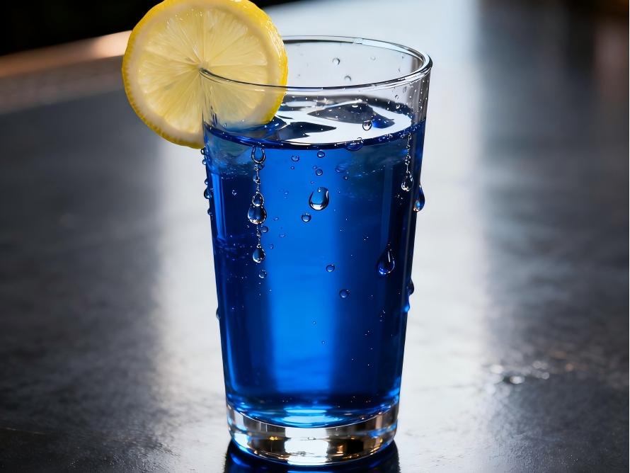
Functional Beverage Breakthrough
· Enabled a sports drink brand to realize its “Ocean Mineral Theme” product concept
· Raw material safety ensures worry-free long-term consumption
· Final product heavy metal levels at just 1/10th of national standard limits
2. Health Supplements: Prioritizing Safety and Efficacy
In health supplements, ingredient purity directly impacts product efficacy and consumer trust:
Protein Nutritional Supplements
· High purity (protein content ≥85%) ensures adequate nutritional content
· Low heavy metal levels meet safety requirements for long-term use
· Helped a renowned brand achieve U.S. FDA registration
Antioxidant Health Supplements
· Preserves intact antioxidant active ingredients
· Safety metrics exceed international pharmacopoeia standards
· Selected as the preferred raw material upgrade for multiple listed companies
3. Cosmetics: Safeguarding Beauty and Safety
As the concept of “cosmetics and food sharing common origins” gains traction, cosmetics demand higher raw material safety standards:
Color Innovation Applications
· Provides safe coloring solutions for premium skincare brands
· Passes cosmetic ingredient safety assessments
· Heavy metal content significantly below limits set by the Cosmetic Safety Technical Specifications
Market Feedback
A product manager from an emerging brand stated: “After using Green Spring Technology's ingredients, our blue mask series not only achieved more stable coloration but, more importantly, completely eliminated concerns about heavy metal migration risks. This has made our products stand out in the competitive landscape.”
4. Enhanced Customer Value
By utilizing our ingredients, clients achieve significant improvements in the following areas:
Trust Value
· End-product inspection pass rate increased to 100%
· Consumer complaint rate decreased by 90%
· Brand reputation significantly enhanced
Innovation Value
· New product development cycle shortened by 50%
· Successful entry into premium markets like infant food
· Product premium potential increased by 20-30%
Compliance Value
· Effortlessly passed domestic and international quality certifications
· 100% customs clearance rate for export products
· Provides robust support for market expansion
“Choosing Green Spring Technology means choosing more than just a raw material—it means choosing peace of mind. Their product quality allows us to focus on market innovation without safety concerns.” — R&D Director, Listed Company
Our raw materials are helping over 200 global enterprises upgrade their products, serving more than 50 product categories cumulatively with zero quality incidents caused by heavy metals.
![]()
Part Three: Partnering with Green Spring Technology to Build a Safe and Pure Future
1. Quality Commitment, Trustworthy
Green Spring Technology is dedicated to providing customers with safe and reliable phycocyanin raw materials. We promise:
· Quality Assurance: Every batch undergoes third-party authoritative testing with comprehensive reports provided
· Stable Supply: Annual production capacity of 200 tons ensures consistent delivery for large-volume orders
· Professional Support: Dedicated technical service team provides comprehensive application guidance
2. Streamlined Collaboration Process
We offer a convenient collaboration pathway:
Step 1: Requirement Discussion
· Outline your specific needs via phone or online consultation
· Our technical consultants will respond within 2 hours
Step 2: Sample Experience
· Free 50g trial sample (includes test report)
· Technical specifications and application recommendations provided simultaneously
Step 3: Customized Solutions
· Tailored solutions based on your product characteristics
· Assistance with formulation testing and process optimization
Step 4: Volume Collaboration
· Formal supply agreement execution
· Establishment of long-term quality tracking system

3. Contact Us
Official Website
https://www.greenspringnatural.com
Professional Consultation
· Email: helen@greenspringbio.com
· Phone: +86 29 88313578
(Weekdays 8:30 AM–5:30 PM)
Instant Communication
· Mobile/WhatsApp: +86 13649243917
(24/7 availability, rapid response)
References
[1] Yao Baozhen. Nutritional evaluation and health-promoting function of spirulina [J]. Food Research and Development, 1998, 6(2): 2.
[2] He Jia, Zhao Qimei, et al. Research on the stability of spirulina phycocyanin [J]. Acta Biologica Sinica, 1998, 15(5): 17.
[3] Claudio S, Mass production of Spirulina [J], Experientia, 1982, 38: 40-43.
-
Prev
Green Spring Technology Solves the Core Challenge: Launches Odorless, Pure Phycocyanin Ingredient
-
Next
Curcumin Ingredient for Next-Gen Functional Foods & Beverages