How to Extract Phycocyanin from Spirulina?
Phycocyanin (PC) is a type of phycobiliprotein, formed by the combination of the blue phycocyanin (phycoerythrin) and soluble protein. Phycocyanin (phycoerythrin) is a type of accessory photosynthetic protein found in cyanobacteria, red algae, cryptophytes and dinoflagellates. comprises a carrier protein and a chromophore auxiliary group (a linearly extended tetrapyrrole compound, see Figure 1) to form a complex protein.
The carrier protein and chromophore are linked by a sulfide bond. Each phycobiliprotein molecule contains two peptide chains, α and β, and each peptide chain contains one or more chromophores covalently bound [1‒2]. The phycobiliprotein molecule contains three chromophores, which are attached to the α-84, β-84 and β-155 positions. The molecular weight of phycobiliprotein is 44 kDa, the isoelectric point (pI) is 4.3, and the maximum absorption wavelength is 620 nm. The purity of phycocyanin is often expressed as A620 nm/A280 nm, and phycocyanin is divided into three types according to purity (P): food grade (P>0.7), reagent grade (0.7<P<3.9), and analytical grade (P>4.0) [3].
Phycocyanin is unstable to light and heat. After 10 days of storage at room temperature under light, the retention rate of the pigment in a 100 mg/L aqueous phycocyanin solution was only 19.34%. After 10 days of storage at 40 °C in the dark, the retention rate of the pigment was only 24.89% [4]. It is thermally stable below 50 °C, but its thermal stability decreases significantly when the temperature reaches or exceeds 60 °C. When the temperature rises to 70 °C, the solution of phycocyanin immediately fades to colorless and a bluish-gray flocculent precipitate appears [5]. Phycocyanin is sensitive to pH. At pH 3 and pH 5, the solubility of phycocyanin is relatively low. At pH 5 to 9, it can better inhibit lipid oxidation, but the emulsifying stability of phycocyanin is better at pH 3 and pH 11[6].
Phycocyanin has functional activities such as anti-tumor, anti-oxidation, anti-inflammation and immunity enhancement. Phycocyanin can inhibit the in vitro migration of lung cancer LTEP-a-2 cells by regulating apoptosis genes [7]. It can enhance the therapeutic effect of radioactive colon cancer by inhibiting the expression of COX-2 [8]. It can significantly increase the SOD enzyme activity in the plasma and liver of mice after radiation, increase the activity of GSH-PX, reduce the content of reactive oxygen species (ROS) in liver tissue, and reduce the oxidative damage caused by radiation to the body [9]. In addition, the protein and chromophore parts of phycocyanin can exert antioxidant effects through different pathways [10]. Phycocyanin can alleviate X-ray-induced pneumonia through the TLR-MyD88-NF-κB signaling pathway[11], promote the proliferation of mouse splenic lymphocytes, and enhance immune activity[12]. Phycocyanin can also inhibit the transformation of osteoblasts into osteoclasts and specific osteoclasts[13]. Phycocyanin is widely used as a natural coloring agent in cosmetics, beverages, ice cream, chewing gum and dairy products [14]. As a functional ingredient, it has attracted widespread attention from the industry.
Phycocyanin can be up to 20% of the dry weight of Spirulina platensis [3,15], which is significantly higher than the 6% of Cyanobacteria from Chaohu Lake [16] and the 7% of Arthrospira maxima [17]. The success of the intensive culture of Spirulina platensis has made it the preferred raw material for the industrial production of phycocyanin. The large-scale extraction, purification and stabilization of phycocyanin has always been the focus of the deep processing of spirulina. This paper reviews the research progress in the extraction, purification and preparation of phycocyanin in spirulina in the past five years, with a view to providing a systematic understanding of the deep development and application of phycocyanin.
1 Research progress in the extraction of phycocyanin
The content of phycocyanin is closely related to the cultivation conditions and processing technology of spirulina. The phycocyanin content of spirulina obtained from different nitrogen sources in the culture medium is different [18], and the phycocyanin content of spirulina irradiated with red light is 42% higher than that of spirulina irradiated with blue light [19]. Spirulina cultivated in spring and summer has a higher phycocyanin content than spirulina cultivated in autumn [20]. Spirulina is commonly dried in several ways: cool drying, sun drying, oven drying, microwave drying, vacuum drying, freeze drying, and spray drying. The drying methods that help to stabilize phycocyanin are freeze drying, cool drying, and spray drying. Other drying methods result in a loss of phycocyanin ranging from 40% to 80% [21]. Phycobiliproteins are intracellular proteins, and the extraction effect is related to the cell wall breaking method and the extraction process parameters.
1.1 Cell wall breaking methods
Common mechanical methods include swelling, repeated freezing and thawing, ultrasonic-assisted cell disruption, high-pressure homogenization, tissue grinding, etc.; there are also chemical solvent methods and biological enzyme methods. Pulsed electric fields and resistance heating methods have also been used in recent years to extract phycocyanin by cell disruption. In practice, several cell disruption methods can be used in combination to achieve the desired effect.
1.1.1 Swelling method
Spirulina dry powder is soaked in an aqueous solution. Due to the difference in osmotic pressure inside and outside the cell, water enters the cell, bursts the cell wall, and the phycobiliprotein is released. The swelling method requires simple equipment and is easy to operate. The disadvantage is that it takes a long time. Yu Jianfeng et al. [22] added spirulina dry powder to a phosphate buffer solution with a pH of 7.0 and allowed it to swell for 6 h. the yield of phycocyanin was 8.08%. MARI et al. [23] soaked dried spirulina powder (liquid-to-powder ratio = 1:250, m:V) in deionized water and phosphate buffer solution (pH 7.0), and measured the average content of phycocyanin in spirulina powder to be 151.80 mg/g.
1.1.2 Repeated freezing and thawing method
The use of a low-temperature freezing environment to freeze a spirulina suspension, followed by thawing at room temperature, can be repeated several times to achieve the effect of cell disruption. The cells are broken and phycobiliproteins are released. The repeated freezing and thawing method is easy to implement, but the disadvantage is that large-scale production takes a long time and is difficult to achieve. Yang Ying [24] dispersed spirulina powder in 0.01 mol/L phosphate buffer (pH 7.0), repeated the freeze-thaw process 3 times, and the purity of the crude extract phycocyanin was 0.97.
1.1.3 Ultrasonic-assisted cell wall disruption method
The main method is to use the cavitation effect of ultrasonic transmission to generate shearing force and shock waves, which fully disrupt the cell wall and release intracellular proteins. The ultrasonic cell wall disruption method has the advantages of a short experimental cycle and high cell crushing rate. However, the disadvantage is that the energy consumption of factory production is high, and the heat generated during the ultrasonic cell wall disruption process causes the temperature of the material to rise, which can easily cause protein denaturation. CHEN et al. [25] used 20 kHz ultrasound for 60 s, with 60 s intervals, at 4 ℃ for 20 min to process spirulina powder solution (liquid-to-solid ratio 1:100, m:V), obtaining a crude extract of phycocyanin with a concentration of 0.73 mg/mL.
1.1.4 High-pressure homogenization
When the material in the high-pressure homogenizer passes through the high-pressure homogenizing valve, the high-speed shearing and impact phenomena generated during the pressurization and sudden decompression process cause immiscible liquid-liquid or liquid-solid experimental materials to form an extremely fine, uniform emulsion. Mari et al. [23] used 1600 bar pressure homogenization to break cell walls, and the content of phycobiliproteins in the crude extract was (291.9±6.7) mg/g.
1.1.5 High-speed shearing method
The strong shearing force generated by the high-speed rotating blade causes the broken material to fully transfer substances with the solvent medium during high-speed flow, promoting the dissolution of soluble substances. Shen Xiangyang et al. [15] dispersed the spirulina platensis at 10,000 r/min and homogenized the mixture for a total of 40 minutes in three batches, the phycocyanin yield was 213.32 mg/g. The mechanical force generated by a colloid mill, ball mill, etc. is used to destroy the cell wall of spirulina. POTT et al. [26] used zirconium oxide beads to continuously grind a fresh spirulina suspension for 48 h, and 90% of the phycocyanin in the Spirulina platensis was dissolved.
1.1.6 Chemical reagent method
Chemical reagents [2-(N-morpholino)ethanesulfonic acid, calcium chloride, etc.] can directly destroy the tissue structure of the cell wall, increase permeability, and cause proteins to flow out of the cell. The treated sample contains less cell impurities, but the introduction of chemical reagents is not conducive to subsequent purification, and chemical reagents are likely to cause damage to the protein structure. PUROHIT, etc. [27] treated spirulina with 2-(N-morpholino)ethanesulfonic acid buffer, and the purity of phycobiliprotein in the crude extract was 0.64. KHAZI, etc. [18] used a 1.5% calcium chloride solution to soak spirulina for 12 h, and the purity of phycobiliprotein reached 1.18.
1.1.7 Biological enzyme method
The cell wall is treated with a biological enzyme to promote the dissolution of intracellular substances. TAVANANDI et al. [28] treated spirulina with 1% lysozyme, and the purity of phycocyanin was 1.19. Ultrasonic-assisted enzymatic treatment (0.6% lysozyme, temperature 37 °C ± 2 °C) is more efficient than the use of surfactants (Triton X-100, Tween 20, Tween 80) and enzymes alone. The extraction efficiency of phycobiliproteins reached 92.73 mg/g, with a purity of 1.09. IZADI et al. [29] treated a spirulina powder suspension with 100 μg/mL lysozyme for 24 h, and the purity of the crude extract of phycocyanin was 0.70, and the protein concentration was 0.23 mg/mL.
1.1.8 Pulse electric field method
Exposing cells to a pulsed electric field causes transmembrane voltage to be formed inside and outside the cell. This causes damage to the cell membrane, which in turn causes the intracellular material to dissolve. AKABERI et al. [30] used a pulsed electric field (40 kV/cm, 1 μs) to treat spirulina cultivated in pH 8 buffer to obtain a crude extract phycobiliprotein purity of 0.51. AOUIR et al. [31] used a pulsed electric field and ultrasound to extract phycobiliproteins. The purity of phycobiliproteins extracted by the pulsed electric field method (P=0.50) was higher than that of the ultrasonic method (P=0.44). The purity of phycobiliproteins in the crude extract was lower than that of the traditional extraction, but the efficiency was higher.
1.1.9 Resistance heating method
A suitable electric field is used to provide resistance through a semiconductor material, which directly generates heat inside the material, causing the membrane to become disordered and produce a polar pattern,which ultimately causes the intracellular components to flow out. PEDRO et al. [32] treated a spirulina powder solution by resistance heating at room temperature and measured the spirulina powder phycocyanin content to be (45.54±1.93) mg/g, which is 51% higher than that obtained by directly heating the spirulina solution to extract phycocyanin.
Hou Zhaoquan et al. [33] compared the freeze-thaw method, the ultrasonic method used alone and the combination of freeze-thaw and ultrasonic, and found that the extraction rate of the combination was 3.07% higher than that of the ultrasonic method used alone. Yu Jianfeng et al. [22] compared the swelling method, the ultra-fine shearing method, the ultrasonic method, the repeated freezing and thawing method, the swelling-ultra-fine shearing method, and the swelling-ultra-fine shearing-ultrasonic method for extracting phycobiliproteins, and found that the swelling-coupled ultra-fine shearing method is suitable for the extraction of phycobiliproteins, and the extraction rate can be as high as 9.22%. In general, the more complete the cell disruption, the higher the phycobiliprotein dissolution rate, but the dissolution of spirulina cell sheath polysaccharides and the like makes subsequent separation and purification of phycobiliproteins more difficult.
1.2 Extraction process
1.2.1 Extraction solvent
Pang Xiaoyu [34] used the freeze-thaw method to compare the effect of 0.3% (m:V) Acolectin-CHAPS (AC) buffer (pH 6.7), 0.1 mol/L phosphate buffer (pH 7.0), and 0.1 mol/L Tris-HCl buffer (pH 7.0) on the extraction of phycobiliproteins. where AC buffer was the most effective, phosphate buffer was second, and Tris-HCl buffer was the least effective. KHAZI et al. [18] used a 1.5% calcium chloride solution to extract phycocyanin, and the purity of the crude phycocyanin extract was 1.18.
1.2.2 Liquid-to-material ratio
Wanida et al. [35] compared the experiments of extracting phycocyanin at three different liquid-to-material ratios of 0.06, 0.04, and 0.02 g/mL. The concentrations of phycobiliproteins in the crude extracts were 6.64, 4.18, and 2.19 mg/mL, respectively. Liu Yuhuan et al. [36] extracted under the conditions of a sodium phosphate-citric acid buffer at pH 7.0 and 30 °C for 1.5 h, and compared the concentrations of spirulina solution at several liquid-to-material ratios of 1:20 to 1:60 (m:V). 30 ℃ conditions, extracted for 1.5 h, compared to the material solution ratio of 1:20~1:60 (m:V) several concentrations of spirulina solution found that, when the material solution ratio is greater than 1:50 (m:V), the amount of phycocyanin (A618 nm) does not increase significantly. A higher liquid-to-material ratio results in a higher concentration of phycobiliproteins in the crude extract, but the yield of phycobiliproteins decreases. A lower liquid-to-material ratio results in more complete protein dissolution and a higher yield of phycobiliproteins, but the subsequent protein concentration and purification work increases.
1.2.3 Ion strength
LI et al. [37] found that the ionic strength of NaCl is greater than 5 g/L to more effectively reduce the chlorophyll extracted at the same time. POTT et al. [26] compared the effect of 0.1–0.8 mol/L calcium chloride solution on the extraction of phycobiliproteins and found that 0.5 mol/L calcium chloride and 0.35 mol/L acetate buffer at pH 6.0 gave the best results.
1.2.4 pH
The polymerization state (monomer, trimer, hexamer or other oligomers) of phycobiliproteins is related to pH. At pH 7.0, 82% of phycobiliproteins exist in the trimer form [38]. Shen Xiangyang et al. [15] compared the effects of different pH (5.0–9.0) buffer systems on the extraction of phycobiliproteins and found that the yield of phycobiliproteins at pH 7.0 can reach 157.75 mg/g.
1.2.5 Temperature
It is common knowledge that proteins are sensitive to temperature. Böcker et al. [5] found by differential scanning calorimetry that phycocyanin undergo rapid depolymerization and denaturation at 50–70 °C, and that phycocyanin trimers are more prone to denaturation than hexamers. WANIDA et al. [39] found by comparison that 0.06 g/mL spirulina swollen in 0.1 mol/L phosphate buffer at 25, 4 and ‒20 ℃ for 12 h, the concentrations of phycocyanin in the crude extracts were 7.52, 6.25 and 4.06 mg/mL, respectively. Increasing the extraction temperature within an appropriate range will help to increase the extraction rate of phycocyanin.
2 Research progress in purification of phycobiliproteins
Spirulina crude extracts contain a wide range of components, including polysaccharides, proteins, mineral salts, and other functional components (chlorophyll, carotene, vitamins, γ-linolenic acid, etc.). The phycocyanins in the crude extracts need to be purified to a certain degree of purity to meet different needs. Common methods of purification of phycobiliproteins include salting-out precipitation, membrane filtration, two-phase extraction, free-flow electrophoresis, column chromatography, etc. The combined use of several purification methods can obtain high-purity phycocyanin.
2.1 Salting-out precipitation method
A low-concentration ammonium sulfate solution (saturation less than 25%) can precipitate impurities such as nucleic acids, chlorophyll, and some miscellaneous proteins, while a high-concentration ammonium sulfate solution (saturation greater than 40%) can precipitate phycobiliproteins. Both methods can be used to precipitate phycocyanin in one step. Zhu Xiaochen [40] used a 40% saturated ammonium sulfate solution for one-step salting out to increase the purity of the crude phycobiliprotein extract from 0.56 to 1.08. Phycobiliprotein can also be purified by using a low-concentration ammonium sulfate solution and a high-concentration ammonium sulfate solution in multiple steps. In the first step, some of the impurities in the crude extract are removed, and in the second step, the phycobiliprotein is collected. Xu Run [41] used 10%/40% saturated ammonium sulfate to increase the purity of phycobiliprotein from 0.59 to 1.62. Shen Xiangyang [42] used 20%/50% saturated ammonium sulfate to increase the purity of phycobiliprotein from 0.3 to 2.3 in two steps.
When purifying phycobiliproteins with ammonium sulfate, the ammonium sulfate introduced into the phycocyanin solution causes problems in subsequent processing.

2.2 Membrane filtration
The membrane filtration process has been used on a large scale in fields such as water treatment, plant extracts, and food processing. Food-grade or higher phycocyanin can be obtained using membrane filtration.
GARCÍA-LÓPEZ et al. [20] used a 0.2 μm microfiltration membrane to filter the crude phycobiliprotein extract, and then used a 10 kDa ultrafiltration membrane to filter it. the purity of phycocyanin increased from 2.65 to 3.72. Qin Song et al. [43] used 300~200 kD and 100~50 kD ultrafiltration membranes to purify phycocyanin concentrate in a stepwise manner, and the purity was >1.0.
2.3 Two-phase solvent extraction method
Qi Qinghua et al. [44] prepared a spirulina powder suspension and used a repeated freeze-thaw method to break the cell wall and add a two-phase solvent (PEG 2000-magnesium sulfate) for extraction. The purity of phycocyanin was increased from 0.78 to 2.64.
Zhu Xiaochen [40] purified phycocyanin by one-step salting-out with ammonium sulfate, and then extracted with a PEG 4000-phosphate double aqueous phase. The purity of phycocyanin was increased from 1.08 to 3.47.
The two-phase extraction method can effectively separate phycocyanin from impurities, but the cost of the two-phase material limits its application in industrial production. In addition, the newly introduced impurities in phycobiliproteins make subsequent separation difficult.
2.4 Free-flowing electrophoresis
Yang Ying [24] used two-step ammonium sulfate precipitation to precipitate the crude phycocyanin extract, and then used free-flow electrophoresis (temperature 14 °C, voltage 500 V, sample flow rate 200 μL/min) to purify phycocyanin, and the purity of phycocyanin was increased from 2.19 to 4.60.
2.5 Column chromatography
Shen Xiangyang [42] used two-step ammonium sulfate precipitation to purify a 2.3% solution of phycocyanin, and then purified the phycocyanin using weak anion exchange chromatography on DEAE Tanrose FF, achieving a maximum purity of 4.0%.
Shao Mingfei [45] added 1.25 mol/L ammonium sulfate to the crude phycocyanin extract for salting out, and then used Macro-Prep Methy1 HIC (methacrylamide ester hydrophobic column chromatography) one-step column chromatography to increase the purity of phycocyanin from 0.506 to 4.017.
Zhang Xiaomeng et al. [46] used a combination of powdered activated carbon and hydroxyapatite column chromatography to increase the purity of phycocyanin from 0.77 to 4.51 using 1.0 mg/mL of crude phycobiliprotein extract.
The column chromatography process has a small capacity and low efficiency, and the green regeneration technology of the resin faces challenges, making it suitable for the production of high-purity phycocyanin.
2.6 A combination of several methods
PUROHIT, etc. [27] used a buffer containing 2-morpholineethanesulfonic acid to swell fresh spirulina. After dialysis of the crude extract, the purity of phycocyanin increased from 0.64 to 1.34, and the purity reached 6.17 after DEAE column chromatography.
Zhang Fayu [16] used repeated freezing and thawing with 1.0 and 1.8 mol/L ammonium sulfate in two steps to increase the purity of the crude phycobiliprotein extract from 0.40 to 1.69. After the two-step salting-out, the phycobiliprotein solution was extracted with a PEG/(NH4)2SO4 aqueuos phase, the purity of phycobiliprotein increased from 1.69 to 2.62; the two-step salt-extracted phycocyanin solution was successively passed through a Cellufine A-500 and HA column, and the purity of phycocyanin could reach 4.59.
3. Research progress in the stabilization of phycocyanin
Phycocyanin is available in liquid phycocyanin, phycocyanin powder, phycocyanin tablets, phycocyanin microcapsules and other preparations. The maintenance of the physiological activity of phycocyanin is closely related to its state of existence. At present, methods for improving the physical and chemical stability of phycocyanin include adjusting the pH, adding stabilizers or preservatives, and preparing phycocyanin microcapsules or nanoparticles.
3.1 Adjusting the pH
Qi Qinghua et al. [44] found that a 0.78% pure phycocyanin solution is more stable when stored at low temperatures, with stability rapidly decreasing after >40 °C. Stability is better at pH 4–7, with the best stability at pH 5. Phycocyanin absorptivity does not change after being stored in the dark for 7 h.
3.2 Adding stabilizers or preservatives
Xu Run et al. [47] found that when the phloroglucinol solution was stored under neutral conditions below 40 °C in the dark, and glucose, sodium chloride and potassium sorbate were added, and then left for 72 h, the preservation rate of phloroglucinol was increased by 53.4%, 31.7% and 35.7%, respectively.
WANIDA et al. [39] compared two solutions: one containing 1 mg/mL phycobiliprotein in 0.4% citric acid and the other without citric acid. After being placed at 80 °C for 1 h, the phycobiliprotein concentration in the solution with citric acid decreased from 65% to 19%, and the concentration of the solution without citric acid decreased from 51% to 11%.

FAIETA et al. [48] studied the effect of sugar on the stability of cyanin by spectrophotometry and circular dichroism, and found that with the extension of storage time, the color of the cyanin solution gradually lost and the structure became unstable. The cyanin solution with sucrose added was more stable, The algin protein is more stable in a 70% sucrose solution than in 20% and 40% sucrose solutions when stored at 65 °C for 1 h.
3.3 Microencapsulation
SCHMATZ et al. [49] used electro-spraying to produce microcapsules of phycocyanin, which protected the activity of the phycocyanin. The PC-PVC (phycocyanin-polyvinyl alcohol) ultrafine particles produced increased the tolerance temperature of the phycocyanin to 216 °C, while the DPPH scavenging rate decreased from 27% for the phycocyanin to 9.2% for the microcapsules.
FAIETA et al. [50] used pure trehalose/trehalose and maltodextrin mixed powder, phycocyanin (0.5% content) to produce phycocyanin microcapsules by freeze-drying and spray drying. It was found that the microcapsules prepared by freeze-drying had an 89% phycocyanin content, while spray-drying had a 77% phycocyanin content. The more trehalose there was, the better the protection of phycocyanin. When phycocyanin microcapsules were placed at 80 °C for 1 h, the phycocyanin content of the lyophilized microcapsules decreased to 64% of the original value, while the phycocyanin content of the spray-dried microcapsules decreased to 90% of the original value.
GUSTININGTYAS et al. [51] used soluble chitosan nanoparticles to prepare microcapsules of phycocyanin. When the mass ratio of chitosan to phycocyanin was 1:0.75, the microcapsules of phycocyanin could be stored at 50 °C for 90 min, and the light absorption at 620 nm remained basically unchanged.
3.4 Chemical modification
MUNAWAROH et al. [52] modified phycobiliproteins with formaldehyde. The maximum absorption wavelength of the phycobiliprotein-formaldehyde complex was shifted to 611 nm. After being exposed to yellow light for 5 h, the light absorption of the phycobiliprotein-formaldehyde complex at 612 nm decreased by 3.95%, and the light absorption of phycobiliprotein at 620 nm decreased by 5.71%. Formaldehyde-modified phycocyanin is more stable than unmodified phycocyanin; however, it is not stable under white light or UV-A (320–400 nm).
Ou et al. [53] modified phycocyanin with polyethylene glycol (PEG). When the molar ratio of PEG to phycocyanin was 5, the modification rate of phycocyanin was 55%. In a simulated experiment in rats, the half-lives of PEG-PC and PC were found to be (1366±55) min and (817±42) min, respectively.
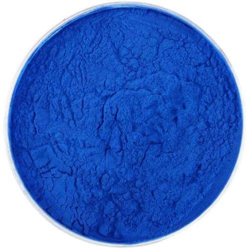
In general, powdered phycocyanin is more stable than liquid phycocyanin, and microencapsulated and chemically modified phycocyanin is even more stable. Currently, phycocyanin generally includes two dosage forms: liquid phycocyanin and powder phycocyanin. Powder phycocyanin is generally produced by spray drying or freeze drying. The main excipients in the product are trehalose, glucose and maltodextrin.

4 Conclusion
In recent years, some progress has been made in the extraction and separation of phycocyanin, but there are still some problems such as low production efficiency and high energy consumption. Research and development is still needed for efficient wall-breaking technology for spirulina and specific purification technology for phycocyanin. The unstable nature of phycocyanin itself limits its application in downstream industries to a certain extent. The technology for stabilizing and maintaining the activity of phycocyanin is still focused on the stability of phycocyanin ingredients. The stability of phycocyanin ingredients after stabilization in food applications still needs to be studied in depth in order to further develop the deep processing and application of phycocyanin.
Reference:
[1] PAN ZK, HU LL. Review on the chemical composition, biological activity and application of Spirulina [J]. Biol Teach, 2020, 45(2): 2–3.
[2] HSIEH LM, CASTILLO G, MOJICA L, et al. Phycocyanin and phycoerythrin: Strategies to improve production yield and chemical stability [J]. Algal Res, 2019, 42: 101600.
[3] KANNAUJIYA VK, SINHA RP. Thermokinetic stability of phycocyanin and phycoerythrin in food-grade preservatives [J]. J Appl Phycol, 2016, 28: 1063–1070.
[4] LV PP, LI CM, YANG DL, et al. Experimental study on the stability of phycocyanin in Spirulina [J]. Guangdong Chem Ind, 2019, 46(5): 60–61.
[5] BÖCKER L, HOSTETTLER T, DIENER M, et al. Time- temperature-resolved functional and structural changes of phycocyanin extracted from Arthrospira platensis/Spirulina [J]. Food Chem, 2020, 316: 126374.
[6] CHEN XH. Effect of pH on the emulsifying activity of C-phycocyanin [J]. Mod Food Sci Technol, 2020, 36(9): 117–125.
[7] YAN Y, HAO S, LI S, et al. In vitro function of phycocyanin on lung cancer LTEP-a-2 cells [J]. J Chin Inst Food Sci Technol, 2018, 18 (8): 24–32.
[8] KEFAYAT A, GHAHREMANI F, SAFAVI A, et al. C-phycocyanin: A natural product with radio sensitizing property for enhancement of colon cancer radiation therapy efficacy through inhibition of COX-2 expression [J]. Sci Report, 2019, 9(1): 19161.
[9] LIU Q, LI WJ, LU LN, et al. Protective effect of phycocyanin on radiation-induced oxidative damage in mice [J]. Nuclear Technol, 2018, 41 (1): 1–6.
[10] MEI X, WANG G, CHENG C, et al. Studies on the antioxidant properties of five different phycocyanins with different purity [J]. Food Sci, 2020,
DOI: 10.7506/spkx1002-6630-20200209-059.
[11] LIU Q, LI W J, LU L, et al. Phycocyanin attenuates X-ray-induced pulmonary inflammation via the TLR2-MyD88-NF-κB signaling pathway [J]. J Oceanol Limnol, 2019, 37(5): 1678–1685.
[12] ZHAO XX, QIU LJ, XUAN CR, et al. Extract of phycocyanin subunits and immunological activity [J]. Chin J Surg Integr Tradit Western Med, 2016, 22(2): 156–160.
[13] MOHAMMED S A, HANAN A, SUNIPA M, et al. C-phycocyanin attenuates RANKL induced osteoclastogenesis and bone resorption in vitro through inhibiting ROS levels, NFATc1 and NF-κB activation [J]. Sci Rep, 2020, 10(1): 2513.
[14] KISSOUDI M, SARAKATSIANOS I, SAMANIDOU V. Isolation and purification of food-grade C-phycocyanin from Arthrospira platensis and its determination in confectionery by HPLC with diode array detection [J]. J Sep Sci, 2018, 41(4): 975−981.
[15] SHEN XY, LIANG X, FU Y, et al. Study of the extraction process of phycocyanin from Spirulina platensis [J]. Mod Food Sci Technol, 2019, 35(7): 198−204, 136.
[16] ZHANG FY. Study on extraction and purification technology of reagent grade phycocyanin and spectral analysis [D]. Hefei: Hefei University of Technology, 2016.
[17] RAY K, MUKHERJEE C. An improved method for extraction and quantification of polyphosphate granules from microbial cells [J]. Protocol Exchange, 2015, 7(1): 67.
[18] KHAZI MI, DEMIREL Z, DALAY MC. Evaluation of growth and phycobiliprotein composition of Cyanobacteria isolates cultivated in different nitrogen sources [J]. J Appl Phycol, 2018, 30: 1513−1523.
[19] WICAKSONO HA, SATYANTINI WH, MASITHAH ED. The spectrum of light and nutrients required to increase the production of phycocyanin Spirulina platensis [J]. IOP Conf Series Earth Environ Sci, 2019, 236,
DOI: 10.1088/1755-1315/236/1/012008.
[20] GARCÍA-LÓPEZ DA, OLGUÍN EJ, GONZÁLEZ-PORTELA RE, et al. A novel two-phase bioprocess for the production of Arthrospira (Spirulina) maxima LJGR1 at pilot plant scale during different seasons and for phycocyanin induction under controlled conditions [J]. Bioresource Technol, 2020, 298: 122548.
[21] NOURI E, ABBASI H, RAHIMI E. Effects of processing on stability of water- and fat-soluble vitamins, pigments (C-phycocyanin, carotenoids, chlorophylls) and colour characteristics of Spirulina platensis [J]. Qual Assur Saf Crop, 2018, 10(4): 335−349.
[22] YU JF, FU J, MA X, et al. Comparison of cell disruption methods for the extraction of phycocyanin from Spirulina platensis [J]. Food Mach, 2017, 55(5): 173−177.
[23] MARI CR, MARJORIE J, ELENA M. Rapid green extractions of C-phycocyanin from Arthrospira maxima for functional applications [J]. JAppl Phycol, 2019, 9(10), 1987.
[24] YANG Y. Methodological research about fast separation and purification of phycocyanin via free flow electrophoresis [D]. Guangzhou: South China University of Technology, 2016.
[25] CHEN KH, WANG SS, SHOW PL, et al. A rapid and efficient technique for direct extraction of C-phycocyanin from highly turbid Spirulina platensis algae using hydrophobic interaction chromatography in stirred fluidized bed [J]. Biochem Eng J, 2018, 140: 47−56.
[26] POTT RWM. The release of the blue biological pigment C-phycocyanin through calcium-aided cytolysis of live Spirulina sp. [J]. Color Technol, 2018, 135(1): 17−21.
[27] PUROHIT A, KUMAR V, CHOWNK M, et al. Processing-independent extracellular production of high purity C-phycocyanin from Spirulina platensis [J]. ACS Biomater Sci Eng, 2019, 5(7): 3237−3245.
[28] TAVANANDI HA, RAGHAVARAO KSMS. Ultrasound-assisted enzymatic extraction of natural food colorant C-phycocyanin from dry biomass of Arthrospira platensis [J]. LWT-Food Sci Technol, 2020, 118:
108802.
[29] IZADI M, FAZILATI M. Extraction and purification of phycocyanin from Spirulina platensis and evaluating its antioxidant and anti- inflammatory activity [J]. Asian J Green Chem, 2018, 2(4): 364–379.
[30] AKABERI S, KRUST D, MÜLLER G, et al. Impact of incubation conditions on protein and C-phycocyanin recovery from Arthrospira platensis post-pulsed electric field treatment [J]. Bioresource Technol, 2020, 306: 123099.
[31] AOUIR A, AMIALI M, KIRILOVA-GACHOVSKA T, et al. The effect of pulsed electric field (PEF) and ultrasound (US) technologies on the extraction of phycobiliproteins from Arthrospira platensis [C]//. Ottawa: Canada IEEE International Humanitarian Technology Conference, 2015.
[32] PEDRO FS, RAFAELA N, CRISTINA MR. Influence of thermal and electrical effects of ohmic heating on C-phycocyanin properties and biocompounds recovery from Spirulina platensis [J]. LWT-Food Sci Technol, 2020, 128: 109491.
[33] HOU ZQ, LIU XY, SHI C, et al. The research of extraction procedure by using method of freezing-thawing and ultrasonic broken frocess to extract phycocyanin from Spirulina algae [J]. J Inner Mongolia Agric Univ (Nat Sci Ed), 2017, 38(2): 69−75.
[34] PANG XY. Study and comparison of the extraction methods of phycocyanin and it,s applicationin remote sensing [D]. Xi'an: Northwest University, 2013.
[35] WANIDA PU, WAREERAT K, SIRILUCK I. Extraction of C-phycocyanin from Arthrospira (Spirulina) and its thermal stability with citric acid [J]. J Appl Phycol, 2017, 30: 231−242.
[36] LIU YH, LI CX, LI DL. Extraction technology and stability of phycocyanin from Spirulina maxima by vacuum freeze-drying [J]. Food Nutr Chin, 2016, 22(9): 51−55.
[37] LI Y, ZHANG Z, PACIULLI M, et al. Extraction of phycocyanin—A natural blue colorant from dried spirulina biomass: Influence of processing parameters and extraction techniques [J]. J Food Sci, 2020, 85(3): 727−735.
[38] AFTARI RV, REZAEI K, MORTAZAVI A, et al. The optimized concentration and purity of Spirulina platensis C-phycocyanin: A comparative study on microwave-assisted and ultrasound‐assisted extraction methods [J]. J Food Proc Preserv, 2016, 39(6): 3080−3091.
[39] WANIDA PU, SIRILUCK I. Physical extraction and extrusion entrapment of C-phycocyanin from Arthrospira platensis [J]. J King Saud Univ Sci, 2019, 31: 1535–1542.
[40] ZHU XC. Preparation of C-phycocyanin and Polysaccharides from Spirulina platensis and their biological activities [D]. Yantai: Yantai University, 2018.
[41] XU R. Research of the storage stability of phycocyanin from Spirulina platensis [D]. Tianjin: Journal of Tianjin University of Science & Technology, 2017.
[42] SHEN XY. Study on extraction and purification of phycocyanin from Spirulina platensis and it,s application [D]. Nanning: Guangxi University, 2019.
[43] QIN S, LIU B, YAN MY, et al. The way of produce a food grade phycocyanin: China, CN 101942014 A [P]. 2011-01-12.
[44] QI QH, CHEN YX, YE Z. The extraction of C-phycocyanin and polysaccharides from Spirulina using aqueous two-phase system [J]. Wuyi Sci J, 2015, 31: 154–160.
[45] SHAO MF. Study on extraction and purification of phycocyanin and pilot preparation technology of food grade phycocyanin from Arthrospira (Spirulina) platensis [D]. Nanjing: Nanjing Agricultural University, 2013.
[46] ZHANG XM, ZHANG FY, WANG JQ, et al. Extraction and purification of C-phycocyanin from blue algae by combined use of powdered activated carbon treatment and column chromatography [J]. J Anhui Agric Univ, 2020, 47(4): 554–558.
[47] XU R, CHEN Y, SUN P. The research of the storage stability of phycocyanin from Spirulina platensis [J]. Food Res Dev, 2017, 38(12): 25–30.
[48] FAIETA M, NERI L, SACCHETTI G, et al. Role of saccharides on thermal stability of phycocyanin in aqueous solutions [J]. Food Res Int, 2020, 132: 1–9.
[49] SCHMATZ DA, MASTRANTONIO DJDS, COSTA JVA. Encapsulation of phycocyanin by electrospraying: A promising approach for the protection of sensitive compounds [J]. Food Bioprod Proc, 2020, 119: 206–215.
[50] FAIETA M, CORRADINI MG, MICHELE AD, et al. Effect of encapsulation process on technological functionality and stability of Spirulina platensis extract [J]. Food Biophy, 2020, 15(1): 50–63.
[51] GUSTININGTYAS A, SETYANINGSIH I, HARDININGTYAS SD, et al. Improvement stability of phycocyanin from Spirulina platensis encapsulated by water soluble chitosan nanoparticles [J]. IOP Conf Seri, 2020, 414(1): 012005.
[52] MUNAWAROH HSH, GUMILAR GG, SHOW PL. Photostabilization of phycocyanin from Spirulina platensis modified by formaldehyde [J]. Proc Biochem, 2020, 94: 297–304.
[53] OU Y, GAO B, MA P. Preparation and pharmaceutical application of modified phycocyanin by polyethylene glycol: China, CN 109535247 A [P]. 2019-03-29.


 English
 French
 Spanish
 Russian
 Korean
 Japanese


