What Is Astaxanthin?

Jan 15,2025
Category:Product News

Astaxanthin (AST) 3, 3'—dihydroxy-4, 4 '—dione—β, β'—carotene, is a carotenoid keto-type oxygen derivative with the chemical formula C40H52O4, which is widely found in various microorganisms and marine animals. The US Food and Drug Administration has approved astaxanthin as a food coloring agent in animal and fish feed [1]. The European Commission considers natural astaxanthin to be a food dye. Natural astaxanthin was first found in shrimp and crab shells, but in very small quantities.


Currently, natural astaxanthin is mainly found in certain algae, yeast and bacteria, with Haematococcus pluvialis being the best source of natural astaxanthin [2]. Haematococcus pluvialis is a green microalga that accumulates high levels of astaxanthin under stress conditions such as high salinity, nitrogen deficiency, high temperature and light [3,4]. Astaxanthin has also been found in plants, a few fungi, chlorococci, chlorella and marine bacteria [5]. Chemically synthesized astaxanthin is readily available and is used as a feed additive for farmed aquatic animals such as fish and shrimp to improve the quality of aquaculture products [6]. The beneficial effects of astaxanthin on aquaculture have been recognized for many years, but research on its potential antioxidant, anti-inflammatory and anti-apoptotic effects on human health has only just begun. This article reviews the detection and biological activity of astaxanthin, with the aim of providing a theoretical basis for the detection and application of astaxanthin in the fields of health food and medicine.


1 In vivo metabolism of astaxanthin

Astaxanthin is absorbed into the body and transported to the liver via the lymphatic system. Its absorption depends on the food components it is ingested with. A high-fat diet can increase the absorption of astaxanthin, while a low-fat diet can reduce it. After ingestion, astaxanthin mixes with bile acids to form micelles in the small intestine. The micelles are partially absorbed by intestinal mucosal cells, integrating astaxanthin into the chyle. The chyle contains astaxanthin, which is released into the lymph during systemic circulation, digested by lipoprotein lipase, and the chyle residue is quickly removed by the liver and other tissues [7]. Among several natural carotenoids, astaxanthin is considered one of the best carotenoids for protecting cells, lipids, and membrane lipoproteins from oxidative damage.


2 Structural characteristics of astaxanthin

The molecular structure of astaxanthin is similar to that of β-carotene. It belongs to the class of unsaturated compounds known as carotenoids. It has a long chain structure with a conjugated double bond in the middle, flanked by two six-membered rings containing oxygen, with the hydroxyl group at the α position of the carbonyl group of the carbonyl group, forming an α-hydroxy ketone structure. This conjugated structure significantly enhances the electron effect of astaxanthin, making it easier to attract unpaired electrons from free radicals, thus exhibiting strong antioxidant capacity.


Unlike other carotenoids, it has unique chemical properties, molecular structure and light absorption characteristics [8]. Its linear structure of “polar-nonpolar-polar” can be inserted across the membrane into the lipid bilayer of the cell or mitochondrial membrane, so that it can easily intercept active molecular substances at the hydrophilic and hydrophobic surfaces of the membrane to exert a powerful antioxidant effect [9].


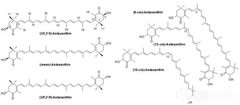
Astaxanthin has two chiral centers, and each of the two chiral carbon atoms can exist in the form of R or S, forming three stereoisomers: 3S, 3S, 3S, 3R and 3R, 3R. The groups connected to the C=C double bond of astaxanthin can be arranged in different ways, divided into cis configuration (Z) and trans configuration (E). The all-trans is the most thermodynamically stable form of astaxanthin. After external energy is applied (e.g. heating), all-trans astaxanthin can undergo cis-trans isomerization, resulting in the appearance of cis-forms at positions 9, 13 and 15. These cis-forms can exist independently or coexist [10]. has one hydroxyl group in its terminal cyclic structure. This free hydroxyl group can form an ester with a fatty acid, which can be either a monoester or a diester. Esterification increases the hydrophobicity, and the diester is more lipophilic than the monoester [6].


3  Standards and testing methods for astaxanthin standards and testing methods

are based on the various structural forms of astaxanthin. In order to further study the relationship between its biological activity and structure, and to facilitate its wider application in medicine, health foods, food additives, aquaculture and cosmetics, it is necessary to establish a simple, rapid and accurate method for analyzing astaxanthin.


The main methods for testing There are two main methods for testing astaxanthin: ultraviolet spectrophotometry and high-performance liquid chromatography. There are also laser Raman spectroscopy, liquid chromatography-mass spectrometry, and thin-layer scanning detection. Ultraviolet spectrophotometry is an early detection method that can only detect the total astaxanthin content and cannot detect the content of various isomers. It is suitable for rapid screening. In high-performance liquid chromatography or mass spectrometry, the extraction method is the key to pretreatment. The extraction methods can be divided into solvent extraction, enzymatic hydrolysis, microwave method, supercritical CO2 extraction, etc. [2].


The main sources of astaxanthin is mainly derived from Haematococcus pluvialis, animal sources such as shrimp, crab, fish, chicken, eggs, etc., as well as synthetic sources. Astaxanthin in these raw materials mainly exists in the form of free and esterified forms. Free astaxanthin only needs to be fully extracted to detect the content of different isomers or the total amount; esterified astaxanthin needs to be saponified or enzymatically hydrolyzed into free astaxanthin before testing. At present there are currently four main national and industrial standards: (1) National Standard GB/T 23745-2009, which is used to test the astaxanthin content in feed additives. The main raw material is synthetic astaxanthin, which is detected using the colorimetric method of a spectrophotometer [11]. (2 ) The national standard GB/T 31520-2015 is a standard for specifically testing the astaxanthin content in Haematococcus pluvialis. The astaxanthin in Haematococcus pluvialis mainly exists in the form of astaxanthin esters. Therefore, this method first saponifies the astaxanthin esters to hydrolyze them into a free state, and then uses the external standard method to quantify them on a C30-HPLC.


This method can simultaneously quantify all-trans, 9-cis, and 13-cis astaxanthin [12]. (3) China Chamber of Commerce for Import and Export of Medicines and Health Products, Group Standard T/CCCMHPIE 1.21-2016, applicable to astaxanthin oil obtained from artificially cultivated Haematococcus pluvialis after extraction and refinement. The astaxanthin ester in the raw material The astaxanthin ester in the raw material is enzymatically hydrolyzed by cholesterol esterase to convert it into free astaxanthin for re-testing, and all-trans, 9-cis, and 13-cis astaxanthin can also be detected simultaneously. This method is derived from the United States Pharmacopoeia, and it is used in the corporate standards of companies such as Senmiao and Alfa [13]. (4 ) Entry-Exit Inspection and Quarantine Industry Standard SN/T 2327-2009: This method is mainly used to detect the levels of canthaxanthin and astaxanthin in imported and exported foods of animal origin. The test subjects are yellow croaker, eel, chicken, eggs, duck liver, pig kidney and milk. Since astaxanthin in these raw materials is present in a free state, so the method directly extracts it with acetonitrile, degreases with hexane, concentrates it and then quantifies it using high performance liquid chromatography (HPLC) external standard method [14].


4 Main biological activities of Astaxanthin's main biological activities

4.1 Antioxidant

Excessive reactive oxygen species can react with proteins, lipids and DNA through chain reactions, leading to oxidation of proteins and lipids and damage to DNA. Damage to these biomolecules is associated with various diseases [15,16]. Oxidative stress is caused by a disturbance of the balance between intracellular oxidation and antioxidant reactions, and is an important mediator in disease pathology. Astaxanthin protects against oxidative damage by neutralizing singlet oxygen, scavenging free radicals to prevent chain reactions, inhibiting lipid peroxidation, enhancing immune system function and regulating gene expression [17,18].


4.2 Anti-inflammatory

Inflammation is a complex series of immune responses that act as host defence mechanisms or responses to bodily damage to initiate tissue repair processes [19]. However , excessive or uncontrolled inflammation can be damaging to host cells and tissues. Astaxanthin is a powerful antioxidant that can prevent inflammation in biological systems. In resting cells, the nuclear factor-kappa B (NF-κB) and the inhibitory kappa B (IκB) form a complex and exist in an inactive form in the cytoplasm. When cells cells are stimulated by extracellular signals, the IκB kinase complex is activated, which phosphorylates IκB and exposes the nuclear localization site of NF-κB. Thus , these stimulus-induced IκB processes lead to the transcriptional regulation of inflammatory genes [20,21]. Astaxanthin blocks NF-κB-dependent signal pathways and inhibits the gene expression of downstream inflammatory mediators such as interleukin (IL)-1β, IL-6 and tumor necrosis factor-α (TNF-α) [22,23]. Astaxanthin also exerts an anti-inflammatory effect by inhibiting cyclooxygenase-1 (COX-1) and nitric oxide (NO) in BV2 microglial cells stimulated by lipopolysaccharide. In vivo studies have also shown that astaxanthin can reduce inflammation in tissues and organs [24,25].


4. 3 Anti-apoptosis

Excessive apoptosis is associated with neurodegenerative diseases, ischemic stroke, heart disease, sepsis and multiple organ dysfunction syndrome. There are various ways to control apoptosis [26]. Many key apoptosis proteins are involved in the two main apoptosis pathways, namely the intrinsic apoptosis pathway (mitochondrial pathway) and the extrinsic apoptosis pathway (death receptor pathway) [27]. Astaxanthin can modify some key apoptosis proteins, thereby preventing the occurrence of related diseases [28]. Astaxanthin enhances the phosphorylation of Bcl-2-associated death protein (BAD) by regulating mitogen-activated protein kinase p38, and downregulates the activity of cytochrome c, caspase-3 and caspase-9. It also


5 Application of astaxanthin in disease prevention and protection

5.1 Protective effect of astaxanthin against ischemia/reperfusion injury

Ischemia/reperfusion (I/R) injury refers to the tissue damage caused when blood supply is restored to an organ after a period of ischemia. A period of hypoxia in a specific region leads to a pathological microenvironment, and the subsequent restoration of blood circulation leads to the activation of inflammatory processes and the production of oxidative damage, rather than a return to normal conditions and function. Reperfusion injury leads to the suppression of the body's defence mechanisms, resulting in an imbalance between the surge in reactive oxygen species (ROS) secretion and the inability of reoxygenated cells to handle this free radical load [34].


In this situation, cell death programs are activated, leading to multi-organ failure. Furthermore, limited oxygen availability is associated with activation of inflammatory signals that control the stability of the transcription factor NF-κB [35] [36], as well as with infiltration of various inflammatory cells (neutrophils, t lymphocytes, monocytes/macrophages) in an adaptive immune response [37]. In reperfusion injury, the adhesion of platelets and leukocytes to endothelial cells is enhanced, leading to a procoagulant state and activation of platelets and leukocytes [38]. This activation can induce the further release of proinflammatory cytokines and chemokines (TNF and IL-1β) in reperfused blood [39]. The endothelial damage is aggravated by the excessive hydroxyl radicals, superoxides and peroxynitrite produced by the reaction of NO in reperfused blood with oxygen.


Ke Xiaoxia et al. [40] showed that astaxanthin exerts a protective effect in hypoxia/reoxygenation (H/R) cardiomyocytes through a PI3K/AKT/HMGB1-dependent pathway. Astaxanthin improves H/R-induced myocardial cell damage, as evidenced by the upregulation of cell activity and the decrease in lactate dehydrogenase/creatine kinase isoenzyme MB levels. In addition, astaxanthin can reduce myocardial cell apoptosis, inhibit the release of IL-6/TNF-α, reduce ROS production, increase superoxide dismutase activity and downregulate malondialdehyde levels. In mechanistic studies, astaxanthin activates PI3K/AKT and inhibits HMGB1 expression. Qiu et al. [41] found that astaxanthin has a protective effect against oxidative stress-induced cytotoxicity of renal tubular epithelial cells and I/R-induced renal injury in mice. In vitro, astaxanthin at a concentration of 250 nM can inhibit the decrease in viability of renal tubular epithelial cells induced by 100 μM H2O2. After 14 days of pretreatment with astaxanthin by gavage, mice can significantly prevent the histological damage caused by I/R. Histological results showed that after pretreatment, the histological score, the number of apoptotic cells and the expression of α-smooth muscle actin were significantly reduced. In addition, astaxanthin can significantly reduce oxidative stress and inflammation in kidney tissue.


Gulten D et al. [42] used astaxanthin to treat mice by gavage for 14 days and then performed a liver I/R experiment. The results showed that astaxanthin treatment significantly reduced the conversion of xanthine aldehyde to xanthine oxidase and the level of tissue protein carbonyl after I/R injury. A recent study by Marisol et al. [43] showed that astaxanthin complexes can reduce muscle damage after I/R of the femoral artery. The researchers blocked the blood flow to the femoral artery of mice and then reperfused it. Astaxanthin showed a compensatory effect against the stress damage. Histological examination showed positive labeling of the macrophage markers CD 68 and CD 163, suggesting a remodeling process. At the same time, the high expression of nuclear factor-erythroid 2-related factor 2 (Nrf2) and NADH Quinone Oxidoreductase 1 (NQO1) reflects the reduction of oxidative damage 15 days after reperfusion. In addition, Otsuka et al. [44] found that astaxanthin significantly reduced retinal I/R injury.


In in vitro studies, astaxanthin inhibited cell death and erythropoietin production in a concentration-dependent manner. Yusuke et al. [45] found that astaxanthin inhibited intestinal mucosal damage and opened tight junctions (TJs), while it had no effect on the P-glycoprotein (P-gp ) has no inhibitory effect, and has no effect on P-gp function. (Immunosuppressants similar to P-gp substrates are used after intestinal transplantation.) It is believed that astaxanthin is effective against intestinal I/R injury. Some studies have shown [46] that astaxanthin treatment can improve learning and memory impairment after repeated cerebral I/R, save the number of surviving pyramidal neurons in the CA1 and CA3 regions, and electron microscopy observations show that astaxanthin can also reduce damage to the ultrastructure of neurons. At the same time, the content of malondialdehyde in the hippocampus decreased, and the levels of reduced glutathione and superoxide dismutase increased. The expression levels of cell chromatin c, caspase-3 and Bax were significantly lower than those in the control group, and the expression of B-cell lymphoma-2 (Bcl-2) was increased.


5.2 Neuroprotective effects of astaxanthin

Although different neurodegenerative diseases can have multiple pathogenic factors, they share some common characteristics. Increased levels of ROS in neurons caused by mitochondrial damage and the release of redox-active metals that interact with oxygen lead to neuronal cell death [47]. A prolonged chronic neuroinflammatory response can lead to neuronal damage and neurodegeneration through the persistent accumulation of neurotoxic proinflammatory mediators [46]. The release of proinflammatory mediators, as well as the release of prooxidants, leads to changes in the morphology and function of intracellular organelles, promoting the occurrence and development of neurodegenerative lesions.


Since oxidative damage and increased neuroinflammation are closely related to the pathogenesis of delayed massive neuronal loss in neurodegenerative diseases, the neuroprotective effect of astaxanthin has attracted interest in the combined treatment and prevention of these diseases [48]. The most common neurodegenerative diseases include Alzheimer's disease, Parkinson's disease, Huntington's disease and amyotrophic lateral sclerosis. Studies have shown that rat neuronal cells PC12 treated with 0.1 μM astaxanthin were protected from 30 μM β-amyloid peptide-induced neurotoxicity. This protection was due to inactivation of Caspase 3, Bax, IL-1β and TNF-α, NF-κB and inhibition of ROS production [49]. Other studies have also confirmed the protective effect of astaxanthin on ROS production and calcium regulation disorders induced by amyloid β in primary hippocampal neurons [50], and can significantly reduce the oxidative stress level and cell death of PC12 cells caused by N-methyl-4-phenylpyridinium iodide damage [51].


Recent studies have reported a direct relationship between astaxanthin and the health effects of the human brain and the promotion of neurogenesis [52,53]. The effects of these two processes decrease significantly with age, leading to a decline in cognitive abilities in the elderly. Although the molecular mechanism has not been fully elucidated, astaxanthin promotes neurogenesis in hippocampus-dependent tasks and improves behavioral performance, which may be the main mechanism of astaxanthin on cognitive function and neurodegeneration caused by Alzheimer's disease.


El-Agamy et al. [5 4] investigated the potential role of astaxanthin and found that it can significantly improve the cognitive decline caused by adriamycin as a protective compound. Al-Amin et al. [55] examined the effect of astaxanthin on the expression of antioxidant enzymes in the brain structure of mice. After 1 month of supplementation with 2 mg/kg astaxanthin, the expression of catalase and superoxide dismutase was enhanced, the level of glutathione was reduced, and the levels of malondialdehyde and advanced protein oxidation products were also reduced in brain regions such as the frontal cortex, hippocampus, cerebellum and striatum, indicating a reduction in lipid peroxidation.


In addition, astaxanthin pretreatment can promote neural cell regeneration and increase the gene expression of glial fibrillary acidic protein, microtubule-associated protein 2, brain-derived neurotrophic factor and growth-associated protein 43 [56]. These proteins are involved in brain recovery. For example, glial fibrillary acidic protein plays an important role in the repair process after damage to the central nervous system and is involved in cell communication and the blood-brain barrier [57]. Microtubule-associated protein 2 is involved in microtubule growth and neuronal regeneration; brain-derived neurotrophic factor is involved in the survival, growth and differentiation of newborn neurons, while growth-related egg 43 can upregulate the activation of protein kinase pathways to promote the formation, regeneration and plasticity of neuronal protrusions [58].


5.3 The skin-protecting effect of astaxanthin

Skin ageing involves a reduction in antioxidants, inflammatory responses, DNA damage, and the production of matrix metalloproteinases that degrade collagen and elastin in the dermis [59–61].


5.3.1 Antioxidant

Several studies have shown that oxidative stress plays an important role in skin aging and damage. Chalyk et al. [62] monitored 31 middle-aged volunteers (17 men and 14 women over 40 years of age) who received 4 mg astaxanthin capsules for 4 weeks. Analysis of the residual skin surface composition showed that there was a significant decrease in the levels of corneal cell shedding and microorganisms. Blood markers showed that there was a sustained decrease in plasma malondialdehyde during astaxanthin administration, indicating that astaxanthin has a strong antioxidant effect and can rejuvenate facial skin. Several researchers have demonstrated that astaxanthin activates the Nrf2/HO-1 antioxidant pathway by producing small amounts of ROS [63,64], stimulates the antioxidant defense system to produce more antioxidants, enhances immunity, and regulates heme oxygenase-1 (HO-1) and other oxidative stress response enzymes. These enzymes are not only markers of oxidative stress, but also participate in the regulatory mechanism of cell adaptation to oxidative damage [65].


5.3.2 Anti-inflammatory

It is well known that sustained oxidative stress can lead to chronic inflammation. Ultraviolet irradiation can increase various pro-inflammatory markers in the skin, such as keratinocytes through the release of pro-inflammatory mediators. Yoshihisa et al. [66] showed that astaxanthin alleviates ultraviolet-induced skin damage by reducing the production of reactive nitrogen species and reducing the expression of inflammatory cytokines. Oral astaxanthin also reduces the levels of UV-irradiation-induced nitric oxide and cyclooxygenase-2, as well as the prostaglandin E2 released by keratinocytes. Astaxanthin can inhibit the production of inflammatory mediators by blocking the activation of NF-κB in human keratinocytes, thus providing new application prospects for the control of skin inflammation [67]. Tominaga et al. [68] conducted a randomized, double-blind, parallel group, placebo-controlled study using 65 healthy female subjects . The study found that IL-1 levels in the stratum corneum increased significantly in the placebo and low-dose (6 mg astaxanthin) groups, but not in the high-dose (12 mg astaxanthin) group, suggesting that long-term preventive astaxanthin supplementation may inhibit age-related skin aging.


5.3.3 DNA repair

Ultraviolet (UV) light-induced skin damage is caused by mutations resulting from errors in DNA repair. A literature search revealed that astaxanthin may play a role in altering the kinetics of DNA repair and may also play a role in limiting UV-induced DNA damage [69]. This inhibition of DNA damage also leads to the stimulation of antioxidant and anti-inflammatory activities via the AKT pathway of oxidative stress enzymes to prevent DNA damage, enhance mitochondrial function and DNA repair. It is hypothesized that astaxanthin may play a role in AKT pathway regulation, thereby contributing to genome stability and combating DNA damage [70]. Park et al. [71] showed in a double-blind controlled study that oral administration of 2 mg or 8 mg astaxanthin capsules for 8 weeks (14 subjects in each group) in healthy women significantly reduced DNA damage, inflammation and oxidative stress and enhanced the immune response (increased levels of natural killer cells, T cells, B cells and IL-6).


5.3.4 Other

Skin ageing is characterized by changes in the various structures of the extracellular matrix, such as collagen, glycosaminoglycans and elastin. These changes lead to various signs of ageing such as loss of skin elasticity, dryness, wrinkle formation and delayed wound healing. UV can lead to the formation of ROS, which in turn leads to an upregulation of matrix metalloproteinase formation, ultimately leading to increased degradation of the extracellular matrix. A large body of literature has shown that astaxanthin can inhibit the expression of matrix metalloproteinases in a variety of cells (such as macrophages, dermal fibroblasts and chondrocytes) [72-74]. Astaxanthin can increase the expression of various biomarkers related to wound healing, such as basic fibroblast growth factor and type I collagen α1 [75].


Astaxanthin can improve the skin's transepidermal water loss, skin smoothness, skin age spots, moisture content and elasticity [76-78]. In a single-blind placebo-controlled study of 49 healthy middle-aged women in the United States,  studied the cosmetic effect of adding 4 mg/day astaxanthin to human skin for 6 weeks. The results showed that the experimental group had significant improvements in fine lines/wrinkles and elasticity compared to the initial values of the measured parameters at baseline. Tominaga et al. [80] conducted two human clinical studies. One was an 8-week open-label non-controlled study in 30 healthy female subjects, in which each person orally consumed 6 mg of astaxanthin and 2 mL of astaxanthin from microalgae per day.


The results showed significant improvements in the subjects' skin wrinkles, age spot size, skin texture, and corneal cell layer water content. In another randomized, double-blind, placebo-controlled study, 36 healthy male subjects were given 6 mg of astaxanthin per day for 6 weeks. The results showed a significant reduction in the parameters of wrinkles, epidermal water loss and sebum oil levels, as well as a significant increase in skin elasticity and moisture content of the stratum corneum.


5.4 Gastric protective effect of astaxanthin

Helicobacter pylori is a Gram-negative bacterium that has been considered to be one of the main causative factors of peptic ulcer disease and gastric cancer. In tissues infected with H. pylori, infiltrating inflammatory cells produce ROS, which cause gastric inflammation by producing a variety of mediators. Wang et al. [81] showed that an algal powder rich in astaxanthin inhibited H. pylori colonization and reduced inflammation in the gastric tissue of infected BALB/cA mice. Lipid peroxidation levels in the astaxanthin group were lower than in the control group, and the growth of Helicobacter pylori was also inhibited, reducing the bacterial load in infected cells. The antioxidant and anti-inflammatory effects of astaxanthin may help to inhibit gastric inflammation caused by Helicobacter pylori. Bennedsen et al. [82] found that adding astaxanthin-containing algal cell extract to the feed can reduce the bacterial load and gastric mucosal inflammation in mice infected with Helicobacter pylori.


The T lymphocyte response in Helicobacter pylori-infected mice differed between the astaxanthin-treated group and the untreated group. After astaxanthin treatment, Helicobacter pylori induced a T helper cell type 1 (Th1) response and the release of interferon (IFN)-γ, which was converted to a Th2 response and the release of IL- 4. Andersen et al. [83] investigated the gastric inflammatory markers and ILS (IL-4, IL-6, IL-8, IL-10, IFN-1) in functional dyspepsia patients treated with astaxanthin. In patients with Helicobacter pylori treated with astaxanthin, upregulation of T helper cells (CD4) and downregulation of cytotoxic T cells (CD8) were observed. However, the bacterial load and cytokine levels in infected tissues were not affected by astaxanthin treatment. Due to the antioxidant activity of astaxanthin, further studies should be conducted to investigate whether astaxanthin inhibits ROS-mediated inflammation in Helicobacter pylori-mediated gastric inflammation.


6 Conclusion

As a substance with strong antioxidant properties, astaxanthin has potential protective effects in diseases such as organ ischemia-reperfusion injury, neurodegenerative diseases, skin diseases, and gastrointestinal diseases. Natural astaxanthin has good application prospects and huge market potential in the pharmaceutical and health product industries due to its safety and wide range of biological activities. Although research on astaxanthin in the pharmaceutical field has continued to deepen in recent years, it is still far from sufficient. Future research should focus on the molecular mechanisms, digestion and absorption, metabolic pathways, etc. of astaxanthin in vivo and in vitro models of different conformations. At the same time, a comprehensive set of testing methods should be established for astaxanthin and its products from different sources to provide an effective theoretical basis for market regulation.


Reference

[1]   Pashkow  FJ,  Watumull  DG,  Campbell  CL, et al. Astaxanthin: A novel potential treatment for oxidative stress and inflammation in cardiovascular disease [J]. Am J Cardiol, 2008, 101(10A): 58–68.

[2]   Zhao  XY,  Zhu  HT,  Bi  YP,  et  al.  Research  of  astaxanithin   in  the Haematococcus pluvialis [J]. Food Res Dev, 2016, 37(4): 191–195.

[3]   Sarada  R,  Tripathi  U,  Ravishankar  GA,  et  al.  Influence  of  stress  on astaxanthin production in Haematococcus pluvialis grown under different culture conditions [J]. Process Biochem. 2002, 37(6): 623–627.

[4]   Sarada  R,  Ranga RA, Sandesh BK, et al. Influence of different culture conditions on yield of biomass and value added products in microalgae [J]. Dyn Biochem Proc Biotechnol Mol Biol, 2012, 6(20): 77–85.

[5]   Ambati RR, Phang  SM, Ravi  S, et al. Astaxanthin: Sources, extraction, stability, biological activities and its commercial applications—a review [J]. Mar Drugs, 2014, 12(1): 128–152.

[6]  Li HM, Gao L. Astaxanthin: Chemicalstructure, biological functionsand usage [J]. Fine Chem, 2003, 20(1): 32–37.

[7]   Olson JA. Absorption, transport, and metabolism of carotenoids in humans [J]. Pure Appl Chem, 1994, 66(5): 1011–1016.

[8]   Higuera–Ciapara  I,  Félix–Valenzuela L, Goycoolea FM. Astaxanthin: a review of its chemistry and applications [J]. Crit Rev Food Sci Nutr, 2006, 46(2): 185–196.

[9]   Goto S, Kogure K, Abe K, et al. Efficient radical trapping at the surface and inside the phospholipid membrane is responsible for highly potent antiperoxidative  activity  of  the   carotenoid   astaxanthin   [J].   Biochim Biophys Acta, 2001, 1512(2): 251–258.

[10]  Sun XJ, Zhao S, Li XX, et al. Research progress of astaxanthin [J]. Food Ferment Technol, 2015, 51(5): 62–66.

[11]   GB/T 23745–2009  GB/T 23745–2009 Feed additive–10% astaxanthin [S].

[12]   GB/T 31520–2015 GB/T 31520–2015 Determination of astaxanthin in Haematococcus–High performance liquid chromatography method [S].

[13]   T/CCCMHPIE 1. 21–2016 T/CCCMHPIE 1. 21–2016 Plant extract–Astaxanthin oil [S].

[14]  SN/T 2327–2009 Determination of canthaxanthin and astaxanthin in foods of animal origin food for import and export [S].

[15]   Edge  R, McGarvey D, Truscott T. The carotenoids as anti–oxidants—a review [J]. J Photochem Photobio B: Biology, 1997, 41(3): 189–200.

[16]   Turrin  NP,   Rivest  S.  Molecular  and  cellular  immune  mediators  of neuroprotection [J]. Mol Neurobiol, 2006, 34(3): 221–242.

[17]   Kamath  BS,  Srikanta  BM,  Dharmesh  SM,  et  al.  Ulcer  preventive  and antioxidative properties of astaxanthin from Haematococcus pluvialis [J]. Eur J Pharm, 2008, 590(1–3): 387–395.

[18]   Rao  AR, Sindhuja H, Dharmesh  SM, et al. Effective inhibition of skin cancer,   tyrosinase,   and   antioxidative   properties   by   astaxanthin   and astaxanthin  esters  from  the  green  alga  Haematococcus  pluvialis  [J].  J Agric Food Chem, 2013, 61(16): 3842–3851.

[19]   Brown GC, Neher JJ. Inflammatory neurodegeneration and mechanisms of microglial killing of neurons [J]. Mol Neurobiol, 2010, 41(2–3): 242–247.

[20]   Shao AW, Wu HJ, Chen S, et al. Resveratrol attenuates early brain injury after subarachnoid hemorrhage through inhibition of NF–κB–dependent inflammatory/MMP–9  pathway  [J].  CNS  Neurosci  Ther,  2014,  20(2):182–185.

[21]   Bhuvaneswari  S,  Yogalakshmi  B,  Sreeja  S,  et  al.  Astaxanthin  reduces hepatic  endoplasmic  reticulum  stress  and  nuclear  factor–κB–mediated inflammation in high fructose and high fat diet–fed mice [J]. Cell Stress Chaperones, 2014, 19(2): 183–191.

[22]   Suzuki Y, Ohgami K, Shiratori K, et al. Suppressive effects of astaxanthin against rat endotoxin–induced uveitis by inhibiting the NF–κB signaling pathway [J]. Exp Eye Res, 2006, 82(2): 275–281.

[23]   Speranza  L,  Pesce  M, Patruno A, et al. Astaxanthin treatment reduced oxidative induced pro–inflammatory cytokines secretion in U937: SHP–1 as a novel biological target [J]. Mar Drugs, 2012, 10(4): 890–899.

[24]   Nakano M, Onodera A, Saito E, et al. Effect of astaxanthin in combination with α–tocopherol or ascorbic acid against oxidative damage in diabetic ODS rats [J]. J Nutr Sci Vitaminol, 2008, 54(4): 329–334.

[25]   Ohgami  K,   Shiratori  K,  Kotake  S,  et  al.   Effects  of  astaxanthin  on lipopolysaccharide–induced inflammation in vitro and in vivo [J]. Invest Ophth Vis Sci, 2003, 44(6): 2694–2701.

[26]   Kam  P,  Ferch N. Apoptosis: mechanisms  and  clinical  implications  [J]. Anaesth, 2000, 55(11): 1081–1093.

[27]   Elmore  S.  Apoptosis:  A review  of programmed  cell  death  [J].  Toxicol Pathol, 2007, 35(4): 495–516.

[28]   Zhang L, Wang H. Multiple mechanisms of anti–cancer effects exerted by astaxanthin [J]. Mar Drugs, 2015, 13(7) : 4310–4330.

[29]   Wu H, Niu H,  Shao A, et al. Astaxanthin as a potential neuroprotective agent for neurological diseases [J]. Mar Drugs, 2015, 13(9): 5750–5766.

[30]   Dong LY, Jin J, Lu G, et al. Astaxanthin attenuates the apoptosis of retinal ganglion  cells in  db/db mice by inhibition of oxidative  stress  [J].  Mar Drugs, 2013, 11(3): 960–974.

[31]   Guo  SX, Zhou HL, Huang CL, et al. Astaxanthin attenuates early acute kidney injury  following  severe  burns  in rats by  ameliorating  oxidative stress and mitochondrial–related apoptosis  [J]. Mar Drugs, 2015,  13(4): 2105–2123.

[32]   Zhang XS, Zhang X, Wu Q, et al. Astaxanthin alleviates early brain injury following  subarachnoid  hemorrhage  in  rats:  possible  involvement  of Akt/bad signaling [J]. Mar Drugs, 2014, 12(8): 4291–4310.

[33]   Kavitha K, Kowshik J, Kishore TKK, et al. Astaxanthin inhibits NF–κB and Wnt/β–catenin signaling pathways via inactivation of Erk/MAPK and PI3K/Akt to induce intrinsic apoptosis in a hamster model of oral cancer [J]. Biochimicaet Biophysica Acta, 2013, 1830(10): 4433–4444.

[34]   Park SW, Kim M, Brown KM, et al. Paneth cell–derived IL–17A causes multi–organ dysfunction after hepatic ischemia and reperfusion injury [J]. Hepatology, 2011, 53(5): 1662–1675.

[35]   Eltzschig  HK,  Eckle  T.  Ischemia  and  reperfusion–from  mechanism  to translation [J]. Nat. Med, 2011, 17(11): 1391–1401.

[36]   Yilmaz G, Arumugam TV, Stokes KY, et al. Role of T lymphocytes and interferon–gamma  in  ischemic   stroke   [J].  Circulation,  2006,   113(17):2105–2112.

[37]   Jin R, Yang G, Li G. Inflammatory mechanisms in ischemic stroke: Role of inflammatory cells [J]. J Leukoc Biol, 2010, 87(5): 779–789.

[38]   Wagner DD, Frenette PS. The vessel wall and its interactions [J]. Blood, 2008, 111(11): 5271–5281.

[39]   Michelle LB, Stephen JL, Philip M, et al. Endothelial dysfunction: From molecular   mechanisms   to   measurement, clinical implications, and therapeutic opportunities [J].   Antioxid   Redox Signal, 2008, 10(9):1631–1674.

[40]   Ke XX, Xu WP, Chen ZQ, et al. The protective  effects  and potential mechanisms of astaxanthin on myocardial hypoxia/reoxygenation injury [J]. Chin Med J Res Prac, 2018, 32(2): 19–22, 26.

[41]   Qiu XF, Kai F, Zhao XZ, et al. Protective effects of astaxanthin against ischemia/reperfusion induced renal injury in mice [J]. J Transl Med, 2015, 13(1): 28.

[42]   Curek GD, Cort A, Yucel G, et al. Effect of astaxanthin on hepatocellular injury   following   ischemia/reperfusion   [J].   Toxicol,   2010,   267(1–3): 147–153.

[43]   Zuluaga TM, Choudat L, Aid–Launais R, et al. Astaxanthin complexes to attenuate muscle damage after in vivo femoral ischemia–reperfusion [J]. Mar Drugs, 2019, 17(6), 354.

[44]   Otsuka  T,  Shimazawa  M,  Inoue  Y,  et  al.  Astaxanthin  protects  against retinal damage: evidence from in vivo and in vitro retinal ischemia and reperfusion models [J]. Curr Eye Res, 2016, 41(11): 1465–1472.

[45]   Takizawa Y,  Kitazato T, Kishimoto H, et al. Effects of antioxidants on drug absorption in in vivo intestinal ischemia/reperfusion [J]. Eur J Drug Metab Pharmacokinet, 2011, 35(3–4): 89–95.

[46]   Yan  X,  Qu  ZZ,  Jian  F,  et  al.  The  protective  effect  of  astaxanthin  on learning and memory deficits and oxidative stress in a mouse model of repeated  cerebral  ischemia/reperfusion  [J].  Brain  Res  Bull,  2017,  131:  221–228.

[47]   Uttara   B,    Singh   AV,    Zamboni   P,    et    al.   Oxidative    stress    and neurodegenerative  diseases:  A  review  of  upstream   and   downstream antioxidant therapeutic options [J]. Curr Neuropharm, 2009, 7(1): 65–74.

[48]   Cho  KS,   Shin  M,  Kim  S,  et  al.  Recent  advances  in  studies  on  the therapeutic potential of dietary carotenoids in neurodegenerative diseases [J]. Oxid Med Cell Longev, 2018: 4120458.

[49]   Chang  CH,  Chen  CY,  Chiou  JY,  et  al.  Astaxanthin  secured  apoptotic death of PC12 cells induced by β–amyloid peptide 25–35: Its molecular action targets [J]. J Med Food, 2010, 13(3): 548–556.

[50]   Lobos  P,  Bruna  B,   Cordova  A,   et  al.  Astaxanthin  protects  primary hippocampal neurons against noxious effects of A β–oligomers [J]. Neural Plast, 2016: 3456783.

[51]   Ye   Q,    Huang   B,   Zhang   X,    et   al.   Astaxanthin   protects    against MPP(+)–induced oxidative stress in PC12 cells via the HO–1/NOX2 axis [J]. BMC Neurosci, 2012, 13: 156.

[52]   Grimmig  B,  Kim  SH,  Nash  K,  et  al.  Neuroprotective  mechanisms  of astaxanthin: A potential therapeutic role in preserving cognitive function  in age and neurodegeneration [J]. Gero Sci, 2017, 39(1): 19–32.

[53]   Yook  JS,  Okamoto  M,  Rakwal  R,  et  al.  Astaxanthin  supplementation enhances adult hippocampal neurogenesis and spatial memory in mice [J]. Mol Nutr Food Res, 2016, 60(3): 589–599.

[54]   El–Agamy SA, Abdel–Aziz AK, Wahdan S, et al. Astaxanthin ameliorates doxorubicin–induced cognitive impairment (chemobrain) in experimental rat model: impact on oxidative, inflammatory, and apoptotic machineries [J]. Mol Neurobiol, 2018, 55(7): 5727–5740.

[55]   Al–Amin  MM,  Akhter  S,  Hasan  AT,  et  al.  The  antioxidant  effect  of astaxanthin is higher in young mice than aged: A region specific study on brain [J]. Metab Brain Dis, 2015, 30(5): 1237–1246.

[56]   Wu W, Wang X, Xiang Q, et al. Astaxanthin alleviates brain aging in rats by  attenuating  oxidative  stress  and  increasing  BDNF  levels  [J].  Food Funct, 2014, 5(1): 158–166.

[57]   Cullen DK, Simon CM, LaPlaca MC. Strain rate–dependent induction of reactive      astrogliosis      and      cell      death      in      three–dimensional neuronal–astrocytic co–cultures [J]. Brain Res. 2007, 1158: 103–115.

[58]   Benowitz  LI,  Routtenberg  A.   GAP–43:  An  intrinsic  determinant  of neuronal development and plasticity  [J].  Trends Neurosci,  1997,  20(2):84–91.

[59]   Kammeyer A,  Luiten RM. Oxidation events and skin aging  [J]. Ageing Res Rev, 2015, 21: 16–29.

[60]   Davinelli  S, Bertoglio JC, Polimeni A, et al. Cytoprotective polyphenols against  chronological  skin  aging  and  cutaneous  photodamage  [J].  Curr Pharm Des, 2018, 24(2): 99–105.

[61]   Zouboulis    CC,   Makrantonaki    E.    Clinical    aspects    and    molecular diagnostics of skin aging [J]. Clin Dermatol, 2011, 29(1): 3–14.

[62]   Chalyk NE, Klochkov VA, Bandaletova TY, et al. Continuous astaxanthin  intake  reduces  oxidative  stress  and  reverses  age–related  morphological  changes of residual skin surface components in middle–aged volunteers [J]. Nutr Res, 2017, 48: 40–48.

[63]   Niu  T,  Xuan  R,  Jiang  L,  et  al.  Astaxanthin  Induces  the  Nrf2/HO–1 Antioxidant  Pathway  in  Human  Umbilical  Vein  Endothelial  Cells  by Generating Trace Amounts of ROS  [J]. J Agric Food Chem, 2018, 66: 1551–1559.

[64]   Saw  CL, Yang AY, Guo Y, et al. Astaxanthin and omega–3 fatty acids individually and in combination protect against oxidative stress via the Nrf2–ARE pathway [J]. Food Chem Toxicol, 2013, 62: 869–875.

[65]   Camera E, Mastrofrancesco A, Fabbri C, et al. Astaxanthin, canthaxanthin and beta–carotene differently affect UVA–induced oxidative damage and expression  of  oxidative  stress–responsive  enzymes  [J].  Exp  Dermatol, 2009, 18(3): 222–231.

[66]   Yoshihisa  Y,   Rehman  MU,  Shimizu  T.  Astaxanthin,  a  xanthophyll carotenoid, inhibits ultraviolet–induced apoptosis in keratinocytes [J]. Exp Dermatol, 2014, 23(3): 178–183.

[67]   Terazawa  S,  Nakajima  H,  Shingo  M,  et  al.  Astaxanthin  attenuates  the UVB–induced secretion of prostaglandin E2 and interleukin–8 in human keratinocytes   by   interrupting   MSK1   phosphorylation   in   a   ROS depletion–independent manner [J]. Exp Dermatol, 2012, 21: 11–17.

[68]   Tominaga K, Hongo N, Fujishita M, et al. Protective effects of astaxanthin on skin deterioration [J]. J Clin Biochem Nutr, 2017, 61(1): 33–39.

[69]   Santocono  M,  Zurria  M,  Berrettini  M,  et  al.  Influence  of  astaxanthin, zeaxanthin and lutein on DNA damage and repair in UVA–irradiated cells [J]. J Photochem Photobiol B, 2006, 85(3): 205–215.

[70]   Ko  JC,  Chen  JC,  Wang  TJ,  et  al.  Astaxanthin  down–regulates  Rad51 expression  via   inactivation   of  AKT   kinase   to   enhance   mitomycin C–induced  cytotoxicity  in human non–small  cell  lung  cancer  cells  [J]. Biochem Pharmacol, 2016, 105: 91–100.

[71]   Park JS, Chyun JH, Kim YK, et al. Astaxanthin decreased oxidative stress and inflammation  and  enhanced  immune response  in humans  [J].  Nutr Metab. 2010, 7: 18.

[72]   Kishimoto  Y,  Tani  M,  Uto–Kondo  H,   et  al.  Astaxanthin   suppresses scavenger  receptor  expression  and  matrix  metalloproteinase  activity  in macrophages [J]. Eur J Nutr, 2010, 49(2): 119–126.

[73]   Chen   WP,   Xiong   Y,   Shi   YX,   et   al.   Astaxanthin   reduces   matrix metalloproteinase     expression     in     human     chondrocytes     [J].     Int Immunopharmacol, 2014, 19(1): 174–177.

[74]   Chou  HY,  Lee   C,  Pan  JL,  et  al.  Enriched   astaxanthin  extract  from haematococcus pluvialis  augments  growth  factor  secretions  to  increase cell  proliferation  and  induces  MMP1  degradation  to  enhance  collagen production in human dermal fibroblasts  [J]. Int J Mol Sci, 2016,  17(6):955.

[75]   Meephansan J, Rungjang A, Yingmema W, et al. Effect of astaxanthin on cutaneous wound healing [J]. Clin Cosmet Investig Dermatol, 2017,  10: 259–265.

[76]   Tripathi  D, Jena G.Astaxanthin  intervention  ameliorates cyclophosphamide–induced  oxidative  stress,  DNA  damage  and  early hepatocarcinogenesis in rat: role of Nrf2, p53, p38 and phase–II enzymes [J]. Mutat Res, 2010, 696(1): 69–80.

[77]   Kavitha  K, Thiyagarajan P, Rathna Nandhini J, et al. Chemopreventive effects of diverse dietary phytochemicals against DMBA–induced hamster buccal pouch carcinogenesis   via    the   induction    of   Nrf2–mediated cytoprotective antioxidant, detoxification, and DNA repair enzymes  [J]. Biochimie, 2013, 95(8): 1629–1639.

[78]   Naidoo K, Hanna R, Birch–Machin MA. What is the role of mitochondrial dysfunction in skin photoaging [J]. Exp Derm, 2018, 27(2): 124–128.

[79]   Yamashita E. The effects of a dietary supplement containing astaxanthin on skin condition [J]. Food Style, 2006, 9(9): 91–95.

[80]   Tominaga K, Hongo N, Karato M, et al. Cosmetic benefits of astaxanthin on human subjects [J]. Acta Biochim Pol, 2012, 59(1): 43–47.

[81]   Wang X, Willén R, Wadström T. Astaxanthin–rich algal meal and vitamin C inhibit Helicobacter pylori infection in BALB/cA mice [J]. Antimicrob Agents Chemother, 2000, 44(9): 2452–2457.

[82]   Bennedsen M, Wang X, Willén R, et al. Treatment of H. pylori infected mice with antioxidant astaxanthin reduces gastric inflammation, bacterial load and modulates cytokine release by splenocytes  [J].  Immunol Lett, 1999, 70(3): 185–189.

[83]   Andersen LP, Holck S, Kupcinskas L, et al. Gastric inflammatory markers and  interleukins   in  patients  with   functional   dyspepsia  treated  with astaxanthin [J]. FEMS Immunol Med Microbiol, 2007, 50(2): 244–248.

Follow Us
Back to the List
Prev

Astaxanthin What Is It Good for Aquaculture?

Next

How to Extract Stevioside?

Need More Information, Please Get in Touch.