Study on Synthesis of Ginsenoside
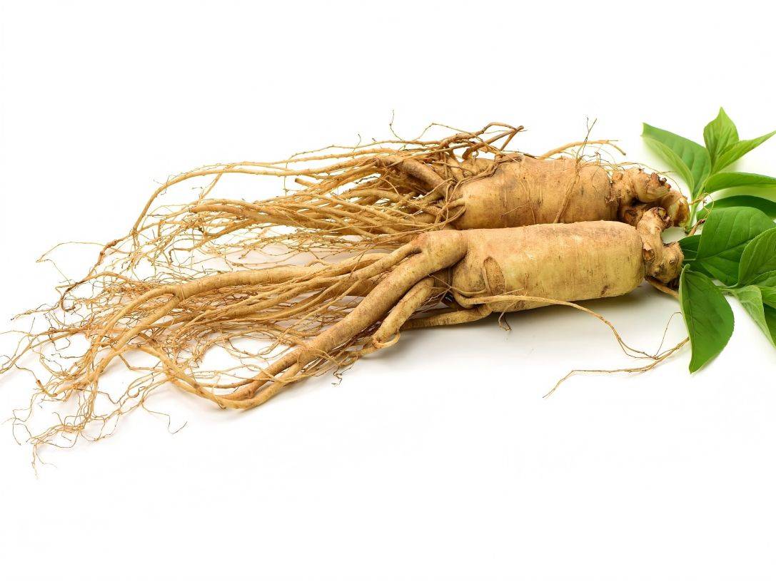
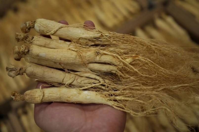
Ginseng (Panax ginseng C. A. Mey), belonging to the genus Panax in the family Araliaceae, is a well-known medicinal plant mainly distributed in northeastern China, Korea and Japan. Ginseng contains various chemical components such as saponins, polysaccharides, polyacetylene and flavonoids. Among them, ginsenoside is a secondary metabolite of ginseng and is its main bioactive component. It has a wide range of physiological and pharmacological activities, including immune system regulation, anti-stress, hypoglycemic, anti-inflammatory, antioxidant and anti-cancer effects. Its mechanism of action is mainly to mobilize the body's internal factors, mobilize neuroprotective mechanisms, and immune mechanisms to exert their effects, with almost no toxicity or adverse reactions.
Ginseng is currently one of the world's best-selling traditional Chinese medicines and is widely used around the world. The total global market consumption of ginseng and related products is estimated to have reached 350 million U.S. dollars [1]. However, ginseng cultivation is very difficult due to the long cultivation time (6–7 years to maturity) and serious plant diseases, such as red skin disease and root rot [1]. Therefore, researchers have studied the tissue and cell cultures of ginseng, such as callus tissue and cell suspensions, to induce root formation in normal roots by Agrobacterium tumefaciens, which in turn produces ginsenosides. However, the production efficiency of ginsenosides by this method is very low. Therefore, metabolic engineering is used to overproduce ginsenosides [2-3], which is an attractive strategy to improve the production efficiency of ginsenosides.
1 Overview of ginsenosides
The main pharmacological active ingredient of ginseng is ginsenoside, which is a triterpene saponin. Ginsenosides are named RX (X = 0, A-1, A-2, B-1, B-2, B-3, C, D, E, F, 20-O-F, G-1, G-2, H-1, ⅆ, X) according to the order of their Rf values from bottom to top on a TLC plate [4]. Ginsenosides are derivatives of sugars, mainly compounds in which the hydroxyl group of a sugar is bound to a non-sugar moiety. The non-sugar moiety is called the aglycone. Ginsenosides are divided into two groups based on the structure of the aglycone: the dammarane type and the oleanane type. The dammarane type is the main type, and its basic skeleton is a tetracycle. According to the position of the sugar groups on carbons 3, 6 and 20, which can be empty or attached to the sugar ring, ginsenosides can be further divided into protoginsenol and protoginsenol. Only one ginsenoside, Ro, is an oleanane-type ginsenoside, with oleanolic acid as the aglycone and a pentacyclic basic skeleton.
At present, ginsenosides have been confirmed to be composed of more than 100 ginsenosides, and more than 40 ginsenosides have been isolated, most of which are damarane-type, including new ginsenosides recently isolated from ginseng buds, processed ginseng, and ginseng leaves. Among them, the most widely studied and noteworthy ginsenosides are Rb1, Rb2, Rc, Rd, Rg1, Rg2, Rg3, Re, Rf, Rh1 and Rh2 [6]. The biological activities of the newly discovered ginsenosides still need to be studied.
2 Biosynthesis of ginsenosides
There are two terpene biosynthetic pathways in plants, namely the MVA pathway and the 2-C-methyl-D-erythritol-4-phosphate (MEP) pathway. Previously, it was generally believed that ginsenosides were synthesized via the mevalonate pathway (MVA pathway) to synthesize IPP and DMAPP, and then 2,3-oxokaurene, which is further modified by hydroxylation and glycosylation to finally produce various ginsenoside monomers. Recent studies have shown that plants can also use the glycolytic intermediates pyruvate and 3-phosphoglycerate as precursors to produce MEP through enzymatic action, and ultimately IPP and DMAPP. Eisen-vaich et al. used a C13 isotope tracer to study the biosynthesis pathway of the anticancer terpene paclitaxel, and the results showed that paclitaxel is mainly synthesized via the MEP pathway [7]. Ginsenosides are also terpenoids, but there are no reports on whether the MEP pathway exists in ginseng.
In plants, the MVA pathway is found to exist in the cytoplasm, while the MEP pathway is found in the plastids [8]. They are separated, but the reaction processes are carried out simultaneously. Although these two pathways exist in two different cellular spaces, they both generate IPP. Whether there is an exchange of IPP between the two pathways and the details of the exchange have always been one of the hot topics in the study of terpene metabolism in plants. Researchers have used inhibitors of key enzymes to inhibit the MVA and MEP pathways separately, confirming that the two pathways are largely independent of each other, while also finding that IPP exchange between the two pathways does occur [9-10]. Therefore, to some extent, the IPP synthesis of the two pathways has a compensatory function, which may also be one of the reasons why the MEP pathway in plants has not been discovered earlier. However, to date, there has been no research on the MEP pathway in ginseng. In addition, it remains to be studied whether both pathways or only one of them plays an important role in ginsenoside synthesis.
In ginseng, the biosynthetic pathways of steroids and triterpenoids share the same precursor, 2,3-oxidosqualene, and the steps of cyclization to form 2,3-oxidosqualene and branching are the same in the two pathways. In ginseng, the synthesis of phytosterols and triterpenoids begins with the product of the 2,3-oxidosqualene cyclization catalyzed by oxidosqualene cyclase (OSCS). In ginseng, β-amyrin synthase (β-AS), dammarane synthase (DS) and cycloartanol synthase (CS) belong to the oxidosqualene cyclase (OSC) family and are located at the branching point of triterpenoid and sterol biosynthesis (Figure 1). CAS catalyzes the formation of cycloartanol, can be used as a precursor for plant sterols. DS and β-AS are both precursors for ginsenosides, with DS providing the tetracyclic dammarane skeleton for the synthesis of dammarane-type ginsenosides and β-AS providing the tetracyclic skeleton for the synthesis of oleanane-type ginsenosides. The intermediates dammarane and β-boswellic acid can be converted into ginsenosides through a series of hydroxylation and glycosylation reactions [11-13]. Cytochrome P450 is thought to be involved in the hydroxylation of the ginsenoside skeleton [14], while glycosyltransferases are involved in the glycosylation of the ginsenoside skeleton.
3 Cloning and research on the genes encoding enzymes involved in ginsenoside biosynthesis
Lee et al. [15] isolated the full-length cDNA clone of SS (PgSS1, accession number: AB115496) through EST analysis of ginseng leaf cDNA libraries. PgSS1 is considered to be a multi-copy gene or a gene with several introns. Overexpression of PgSS1 enhanced the activity of the PgSS1 enzyme, resulting in a significant increase in plant sterol and ginsenoside content. These results indicate that PgSS1 is not only a key regulatory enzyme in phytosterol biosynthesis, but also in ginsenoside biosynthesis. The same results were also found in the heterologous overexpression of Panax ginseng PgSS1 [16], in which the levels of phytosterols (B-sitosterol, stigmasterol) and triterpene saponins increased by 2.0 to 2.5 times in transgenic Panax ginseng. In addition, this suggests that in other plants, heterologous overexpression of genes involved in ginseng triterpene saponin biosynthesis can be used to increase ginsenoside levels and elucidate the biosynthesis mechanism of ginsenosides.
Kushiro et al. [12] isolated two different homologous cDNA clones encoding β-AS synthase (PNY1 and PNY2) from ginseng root hair. These two β-ASs may have a common ancestor that evolved through multiple copies and mutations during evolution. The key internal part of the enzyme that forms β-asarone (PNY1) has been determined. In addition, a site-directed mutation study of PNY1 identified a single amino acid residue, Tyr261, that is critical for product specificity. β-Amyrin and its metabolites are often tissue-specific [17], which may be why only one type of oleanane-type saponin (Ro) has been identified from ginseng.
Dammarane synthase (DS) is considered to be the most important biosynthetic enzyme of ginsenosides. Under its catalytic action, 2,3-oxidosqualene is converted into (20S)-dammarane, not (20R)-dammarane. Recently, researchers have used RT-PCR technology to clone the dammarane-II synthase gene [18]. This DS contains a 2,310 bp ORF encoding a polypeptide of 770 amino acids, and the predicted molecular mass of this polypeptide is 88.3 kDa. In addition, RNA interference DS in transgenic ginseng can silence DS expression, resulting in a 84.5% reduction in saponin production in ginseng roots [19]. These results indicate that DS is a key enzyme involved in ginsenoside biosynthesis, and therefore overexpression of DS can significantly enhance ginsenoside biosynthesis.
So far, only SS, DS, β-AS and CS have been studied. In ginseng, a gene (accession number AB009031) encoding for the production of ginsenoside protopanaxatriol has been identified [20], suggesting a novel plant sterol synthesis pathway in ginseng. In addition, the results of expression sequence tag (EST) analysis of cDNA libraries from different tissues of ginseng [5, 14, 21] showed that candidate genes related to ginsenoside biosynthesis encode enzymes such as HMGR, FRS, geranylgeranyl diphosphate synthase, cytochrome P450, glycosyltransferase, β-glucosidase and lupeol synthase (LUS).
4 Outlook
Among the various chemical components of ginseng, ginsenosides are its main active ingredients. Currently, most studies have focused on the saponin components. Before the MEP pathway was discovered in bacteria and plants, the MVA pathway was considered the only synthetic route for the synthesis of triterpenoid saponins from IPP and DMAPP. The MEP pathway has now been shown to exist in a variety of plants; however, more research is needed on the MEP pathway in ginseng.
Molecular biology and enzymology techniques have been effectively used to reveal the mechanism of ginsenoside biosynthesis, and more and more complete cDNA sequences of genes and candidate genes encoding enzymes related to ginsenoside biosynthesis have been obtained. In addition, EST technology has been extensively used for gene cloning and expression of squalene synthase (HMGR, FPS, farnesyl diphosphate synthase, SE) required for ginsenoside synthesis and enzymes required in subsequent steps (cytochrome P450, glycosyltransferase, B-glucosidase). In addition, the discovery of the candidate gene for the synthesis of lupeol and lanosterol in ginseng has enhanced understanding of the metabolic pathways in ginseng. Traditionally, ginseng roots have been considered the main tissue for ginsenoside biosynthesis. However, the DS responsible for most ginsenoside biosynthesis is expressed at the highest level in ginseng flower buds [19]. This suggests that ginseng flower buds may be an ideal material for further dissecting the ginsenoside biosynthesis pathway.
So far, the main methods used to identify genes encoding enzymes involved in ginsenoside biosynthesis have been RT-PCR [12–13] and EST analysis [5, 14, 21]. A ginseng bacterial artificial chromosome library based on ginseng genomics has been constructed. These resources can be used not only to identify genes related to ginsenosides, but also to elucidate the regulatory mechanisms of gene expression. In recent years, RNAi has become a very effective technical means in plant metabolic engineering. It can effectively inhibit the expression of specific genes and can be used as a tool for the future discovery of genes involved in ginseng metabolic regulation and functional verification. The use of RNAi technology can analyze genes related to ginsenoside synthesis on a large scale and with high efficiency, and can more effectively and accurately identify possible metabolic regulatory genes and verify their function [22]. At present, although substantial progress has been made in revealing the ginsenoside synthesis pathway, research on the catalytic level of the relevant enzymes has not yet been carried out. In addition, the subsequent steps of ginsenoside biosynthesis still need to be clarified, and there is still a long way to go to analyze the biosynthesis of ginsenosides.
Ginseng saponins are an important component of secondary metabolites, and their content and composition are mainly determined by the key enzymes in biosynthesis and their expression levels in cells. The metabolism of plant sterols and triterpenoids is a highly complex and dynamic process regulated by multiple factors. There are still many questions to be answered before the metabolic pathway of ginsenosides can be fully elucidated. However, considering the economic and pharmacological importance of ginseng, this is still an important area worth working on.
Reference:
[1]HONG S G,LEE K H,KWAK J,et al.Diversity of yeasts associated with Panax ginseng[J].Journal of Microbiology,2006,44: 674-679.
[2] Wu Qiong, Zhou Yingqun, Sun Chao, et al. Ginsenoside biosynthesis and secondary metabolism engineering [J]. Chinese Journal of Biological Engineering, 2009, 29(10): 102-108.
[3]LIANG Y,ZHAO S.Progress inunderstanding of ginsenoside biosynthesis [J].Plant Biology,2008,10:415-421.
[4]OKAZAKI H,TAZOE F,OKAZAKI S,et al.Increased cholesterol biosyn- thesis and hypercholesterolemia in mice overexpressing squalene synthase in the liver[J].Journal of Lipid Research,2006,47: 1950-1958.
[5]KIM M K,LEE B S,IN J G,et al.Comparative analysis of exp ressed se- quence tags ( ESTs) of ginseng leaf[J].Plant Cell Rep,2006,25: 599 -606.
[6]HELMS S.Cancer prevention and therapeutics: Panax ginseng[J].Alterna- tive Medicine Review,2004,9: 259-274.
[7]EISENVAICH W,MENHARD B,HYLANDST P J,et al.Studies on the bi- osynthesis of taxol: the taxane carbon skeleton is not of mevalonoid origin [J].Biochemistry,1996,93: 6431-6436.
[8]SEEMANN M,TSE SUM BUI B,WOLFF M,et al.Isoprenoid biosynthesis in plant chloroplasts via the MEP pathway : direct thylakoid /ferredoxinde- pendent photoreduction of GcpE/ IspG[J].FEBS Letters,2006,580: 1547 - 1552.
[9]HEMMERLIN A,HOEFFLER J F,MEYER O,et al.Cross-talk between the cytosolic mevalonate and the plastidial methylerythritol phosphate pathways in tobacco bright yellow-2 cells[J].Journal of Biological Chemistry,2003,278: 26666-26676.
[10]ROHDICH F,ZEPECK F,ADAM P,et al.The deoxyxylulose phosphate pathway of isoprenoid biosynthesis: studies on the mechanisms of the re- actions catalyzed by IspG and IspH protein[J].Proceedings of the Na- tional Academy of Sciences,USA,2003,100: 1586-1591.
[11]KUSHIRO T,OHNO Y,SHIBUYA M,et al.In vitro conversion of 2,3 -oxidosqualene into dammarenediol by Panax ginseng microsomes[J].Biological & Pharmaceutical Bulletin,1997,20: 292-294.
[12]KUSHIRO T,SHIBUYA M,EBIZUKA Y.Beta-Amyrin synthase: Cloning of oxidosqualene cyclase that catalyzes the formation of the most popular triterpene among higher plants[J].Eur J Biochem,1998,256: 238-244.
[13] KUSHIRO T,SHIBUYA M,EBIZUKA Y.Molecular cloning of ox- idosqualene cyclase cDNA from Panax ginseng: the isogene that encodes betaamyrin synthase.Towards natural medicine research in the 21st century[J].Excerpt a Medica International Congress Series,1998,1157 : 421-428.
[14]JUNG J D,HAHM Y,HUR C G,et al.Discovery of genes for ginsenoside biosynthesis by analysis of ginseng exp ressed sequence tags[J].Plant Cell Rep,2003,22: 224-230.
[15]LEE M H,JEONG J H,SEO J W,et al.Enhanced triterpene and phytos- terol biosynthesis in Panax ginseng overexpressing squalene synthase gene[J].Plant Cell Physiol,2004,45 (8) : 976-984.
[16]SEO J W,JEONG J H,SHIN C G,et al.Overexpression of squalene syn- thase in Eleutherococcus senticosus increases phytosterol and triterpene accumulation[J].Phytochemistry,2005,66: 869-877.
[17]PHILLIPS D R,RASBERY J M,BARTEL B,et al.Biosynthetic diversity in plant triterpene cyclization[J].Current Opinion in Plant Biology,2006, 9: 305-314.
[18]PIMPIMON TANSAKUL M S,TETSUO KUSHIRO,YUTAKA EBIZUKA. Dammarenediol-II synthase,the first dedicated enzyme for ginsenoside bi- osynthesis,in Panax ginseng[J].FEBS Letters,2006,580: 5143-5149.
[19]HAN J Y,KWON Y S,YANG D C,et al.Expression and RNA interfer- ence-induced silencing of the dammarenediol synthase gene in Panax ginseng[J].Plant Cell Physiol,2006,47 ( 12) : 1653-1662.
[20]SUZUKI M,XIANG T,OHYAMA K,et al.Lanosterol synthase indicotyle- donous plants[J].Plant & Cell Physiology,2006,47: 565-571.
[21]CHOI D W,JUNG J,HA Y I,et al.Analysis of transcripts in methyl jas manate-treated ginseng hairy roots to identify genes involved in the bio- synthesis of ginsenoides and other secondary metabolites[J].Plant Cell Rep,2005,23: 557-566.
[22] Pan Xichun, Sun Min, Zhang Lei, et al. RNA interference and its application in metabolic engineering of medicinal plants [J]. Chinese Herbal Medicine, 2005, 36(9): 1281-1284.


 English
 French
 Spanish
 Russian
 Korean
 Japanese