

 English
 French
 Spanish
 Russian
 Korean
 Japanese
What Is the Use of Hyaluronic Acid Hydrogel in Tissue Repair?
1 Introduction
Hyaluronic acid (HA), also known as hyaluronic acid, is a negatively charged linear glycosaminoglycan formed by alternately linking glucuronic acid and glucosamine as disaccharide units [1]. As a major component of the extracellular matrix, hyaluronic acid is found in almost all mammalian tissues [2]. Hyaluronic acid is mainly recognised by receptors such as CD44 and RHAMM, which activate intracellular signalling pathways involved in inflammatory responses and tissue regeneration [3]. Hyaluronic acid of different molecular weights has different functions.
Low molecular weight hyaluronic acid plays a pro-angiogenic and pro-inflammatory role: the promotion of angiogenesis is related to the activation of the MAPK/ERK signalling pathway, which leads to the activation of ERK1/2 and the increase of endothelial cell migration; the pro-inflammatory role is due to the fact that low molecular weight hyaluronic acid participates in the recognition of the TLR of the innate immune response by endogenous danger signals, and it firstly binds to the TLR receptor, triggering the signalling cascade reaction and thus the production of pro-inflammatory cytokines.
It triggers the production of pro-inflammatory cytokines and chemokines, and then activates macrophages and induces the maturation of dendritic cells, thus exerting a pro-inflammatory effect [4-5]. High molecular weight hyaluronic acid has both angiogenic and anti-inflammatory properties: angiogenesis is inhibited by inhibiting the expression of endothelial cell early response genes (e.g., c-fos, c-jun, and Krox-20)[6] ; anti-inflammatory effects are due to the fact that high molecular weight hyaluronic acid acts as an inhibitor of inflammation to inhibit TLR2 signalling, and the thin layer of structure it forms recruits the inflammatory cells to an inactive state thereby inhibiting the overall inflammatory process. Its thin layer structure recruits inflammatory cells and keeps them inactive, thus inhibiting the whole inflammatory process [7].
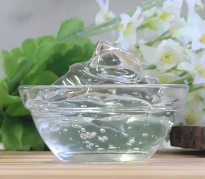
Hydrogels are a class of tissue-engineered products with a three-dimensional network-like structure, whose internal pores allow the entry and adhesion of living cells, as well as the exchange of gases, nutrients and metabolites, and are suitable for the repair of tissue damage and the regulation of cell behaviour [8]. Hyaluronic acid hydrogels can be prepared from natural polymers, synthetic polymers, or complexes of the two according to the actual needs, while hyaluronic acid hydrogels have been widely used in the field of tissue engineering due to their good biocompatibility, bioactivity, and modifiability [9].
Hyaluronic acid hydrogels are widely used in tissue engineering due to their biocompatibility, bioactivity and modifiability [9], and the carboxyl group of hyaluronic acid molecules can be fully ionised at physiological pH, which makes them highly hydrophilic and water-retentive, and allows the formation of mucoadhesive hydrogels even at low concentrations [1]. In recent years, tissue repair using hyaluronic acid hydrogel as scaffold material is often combined with exogenous stem cell transplantation and tissue microenvironment regulation to fully simulate the physiological environment and mobilise the regenerative function of the organism, which brings a ray of hope for the healing of tissue injuries, and this paper focuses on the application prospects of hyaluronic acid hydrogel in damaged skin repair, bone repair, cartilage repair and central nervous system repair.
2 Hyaluronic acid hydrogel and skin repair
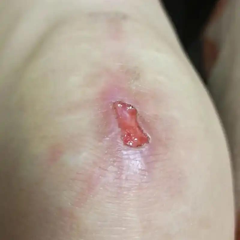
The skin is capable of healing itself in the case of minor trauma, but when the trauma exceeds the compensatory effect of the skin, it is necessary to intervene in a timely manner, especially to close the wound, otherwise it may lead to skin infection, excessive inflammation and complications, and even threaten the patient's life [10]. The wound healing process is divided into four phases: haemostasis, inflammation, proliferation and remodelling, which involves multiple aspects and is a coordinated process of cell proliferation, angiogenesis and extracellular matrix deposition. Hyaluronic acid is a natural polysaccharide produced by fibroblasts during the proliferative phase of wound healing, which can mediate cellular signalling to promote cell migration [11]. Hyaluronic acid-based hydrogels not only provide a moist and relatively closed microenvironment, but also help collagen deposition, granulation tissue and new blood vessel formation, and promote rapid re-epithelialisation of the skin, making them ideal dressings for skin wounds [12].
Hyaluronic acid has been widely used in the preparation of hydrogels with different functions to meet the needs of different types of skin wounds, especially with the addition of antibacterial, anti-inflammatory, pro-angiogenic and other active ingredients, which can more effectively promote wound healing. Antimicrobial hyaluronic acid hydrogels can be prepared by combining antibacterial natural polysaccharides such as chitosan on the one hand, and antibacterial active ingredients such as nanosilver on the other hand [13-14]. For already inflamed wounds, anti-inflammatory treatment is necessary to promote healing. Hyaluronic acid grafted with β-cyclodextrin can form a new type of self-healing hydrogel with adamantane polyethylene glycol through subject-object interaction.
At the same time, the hydrophobic cavity of β-cyclodextrin can carry the hydrophobic anti-inflammatory drug dexamethasone to inhibit inflammation [15]. Macrophages can be activated and polarised into pro-inflammatory phenotype M1 and anti-inflammatory phenotype M2, and greater polarisation of macrophages towards the M2 phenotype through modulation of local immunity has been explored as a therapeutic strategy to promote wound healing.Saleh B et al [16] synthesised nanogels to encapsulate miR-223 mimics through electrostatic interactions between hyaluronic acid-polyethyleneimine and hyaluronic acid-polyethylene glycol. Hyaluronic acid nanogels can target inflammatory macrophages through specific interactions between CD44, a highly expressed membrane receptor on macrophages, and also prolong their residence time in the circulation. Tiny RNA miR-223 encapsulated in hyaluronic acid nanogels can reprogramme macrophages to fight inflammation and promote wound healing. Oligohyaluronic acid can stimulate the secretion of vascular endothelial growth factor (VEGF) to trigger the formation of new blood vessels, and Wang et al. [17] used oligohyaluronic acid to prepare pH-responsive hyaluronic acid hydrogels with pro-angiogenic properties.
3 Hyaluronic acid hydrogels and bone repair
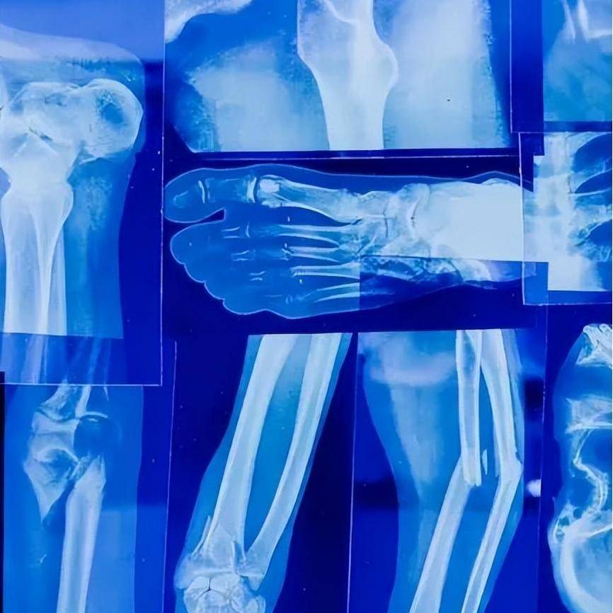
The mainstay of clinical bone defect treatment is the implantation of autologous bone or bone substitutes, which are associated with many risks such as infection and immune rejection [18]. Ideally, bone substitutes should be made of biocompatible materials that mimic the structure, characteristics, and function of natural bone, and 3D bioprinting is an ideal method for preparing biomimetic hydrogels. Cell-loaded hydrogel scaffolds prepared with methacrylated gelatin and hyaluronic acid as printing inks were able to maintain the integrity of the scaffold network and significantly promote the formation of bone matrix after 28 d of incubation in mineralisation-inducing medium. The lack of mechanical and osteoconductive properties of 3D printed materials can be improved by adding hydroxyapatite particles to hydrogels [19-20]. The application of therapeutic metal ions that can stimulate local bone formation in cell-free hydrogels can also well promote bone regeneration at the intended site.
Zhang et al [21] prepared a nanocomposite hydrogel based on hyaluronic acid and self-assembled bisphosphonate-magnesium nanoparticles, which not only enhanced the network structure of the hydrogel by using nanoparticles with acrylate groups on the surface as an effective multivalent cross-linking agent, but also promoted the mineralisation of the hydrogel and mediated the sustained release of Mg2+ . A Wnt5a mimetic hexapeptide (Foxy5 peptide) bound to hyaluronic acid hydrogels mimics the bone-enhancing microenvironment in trabeculae by activating non-classical Wnt signalling, enabling MSCs to ‘sense’ mechanical forces and promote osteogenesis [22]. Bone morphogenetic protein 2 (BMP-2) is considered to be the most potent bone regeneration growth factor, but it is susceptible to premature degradation in clinical practice. Hyaluronic acid has a carboxyl group, and by adjusting the protonation state of hyaluronic acid carboxylic acid residues in covalently crosslinked hydrogels, the molecular interactions with BMP-2 can be utilised to achieve the intelligent release of BMP-2 at physiological pH values [23].
4 Hyaluronic acid hydrogel and cartilage repair
Once bone disease involves joints, cartilage damage inevitably occurs. The regeneration and repair of articular cartilage, which has a poor self-repairing ability, is extremely challenging, and a good clinical solution is the application of exogenous umbilical cord blood-derived mesenchymal stem cells (MSCs) [24]. Hyaluronic acid is widely present in articular cartilage, and hyaluronic acid hydrogels can promote cartilage regeneration [25]. Hyaluronic acid hydrogels for cartilage repair are subjected to mechanical loads caused by patient's exercise for a long period of time, and therefore, their mechanical properties are very demanding.The micellar crosslinked hyaluronic acid hydrogels prepared by Ren et al [26] have excellent stiffness and toughness, and they are promising materials for cartilage repair.The hydrogels obtained by Kim et al [27] by adding nano-clay to bis-phosphonate modified hyaluronic acid also have excellent mechanical properties.The hydrogels obtained by Kim et al [27] have excellent mechanical properties. Kim et al. [27] obtained a hydrogel by adding nanoclay to bisphosphonate-modified hyaluronic acid.
Rheumatoid arthritis, tumours and other joint diseases may cause damage to osteochondral bone. The complex repair process of osteochondral cartilage requires the addition of other active ingredients to hyaluronic acid hydrogels, and Yang et al [28] used hyaluronic acid hydrogels combined with Icariin to promote not only cartilage and osteogenesis, but also the repair of the calcified layer in vitro. As cartilage and subchondral bone have significant differences in chemical composition and biological profile, Liu et al. [29] prepared a biomimetic biphasic osteochondral scaffold that can promote the repair of cartilage and subchondral bone, respectively. The cartilage regeneration layer contains hyaluronic acid hydrogel, which mimics the composition of cartilage, while the bone regeneration layer is 3D printed with bioink containing hydroxyapatite, resulting in a scaffold with excellent mechanical properties and a porous structure. The cartilage and bone regeneration layers were supplemented with different inducers to regulate the differentiation of MSCs into chondrocytes and osteoblasts, respectively. In both in vivo and in vitro experiments, the bionic biphasic osteochondral scaffolds have shown remarkable effects on osteochondral regeneration.
5 Hyaluronic acid hydrogel and central nervous system repair
The central nervous system (CNS), including the spinal cord and the brain, is difficult to regenerate after injury due to microenvironmental inhibition and glial scarring [30]. Hyaluronic acid hydrogel can mimic the extracellular matrix of natural nerve tissue and bridge the injury site, which is beneficial to CNS injury repair. At the same time, loading the adhesion peptide PPFLMLLKGSTR in hyaluronic acid hydrogel can significantly promote the adhesion growth of MSCs, and play the role of MSCs in compensating for the damaged neurons and neurotrophins, thus achieving more effective spinal cord tissue repair [31]. Neural progenitor cells in the subventricular region proliferate in large numbers after a stroke, but they do not migrate to the stroke site, which means that recruiting endogenous neural progenitor cells to the lesion site is a more effective way than injecting exogenous stem cells directly via hydrogel.
The hyaluronic acid injectable particle hydrogel prepared by Nih et al [32] can deliver nerve growth factor through hyaluronic acid, and at the same time, the particles are annealed to each other in situ to form a microporous monolithic scaffold, which can mediate the rapid migration of endogenous neural progenitor cells, and the synergistic effect of nerve growth factor and neural progenitor cells in the focal site will lead to the repair of the brain injury. Angiogenesis in brain repair can change the inhibitory microenvironment after traumatic brain injury. Lu et al[33] used a hydrogel obtained by modifying hyaluronic acid with a VEGF mimetic peptide to repair brain injury by promoting angiogenesis and inhibiting the formation of glial scar tissue.
6 Summary and Outlook
Hyaluronic acid hydrogels are widely used in tissue engineering, and the modification of their biomimetic structure and the loading of bioactive components can make the structure and function of the hydrogel materials meet the requirements of various types of tissue trauma treatment. Hyaluronic acid hydrogels can not only work well with stem cells for repair, but can also be used for targeted therapies due to its receptor-specific binding properties. However, hyaluronic acid functions differently depending on its molecular weight, and therefore, the impact of functional changes as the implanted hydrogel degrades to lower molecular weights needs to be considered.

The application of hyaluronic acid hydrogel in various types of tissue repair has been a hot research topic, and the design of hydrogel can meet different needs and improve the therapeutic efficiency. (1) The physical properties of hyaluronic acid hydrogels, such as softness and hardness, can regulate the behaviour of the cells encapsulated in them, and the cells will improve the regulation of the hydrogel by secreting proteins and other means to adapt to the needs [34-35]. Therefore, researchers can explore the interaction between hyaluronic acid hydrogels and cells to better regulate cellular behaviour and promote tissue repair. (2) The wound healing process is very complex, and a multifunctional smart hydrogel system that can rapidly heal wounds through more than one biological mechanism is more in line with clinical needs and an important direction for wound dressing research. (3) Nanoparticles have irreplaceable advantages in the field of drug loading, combined with the existing research on hyaluronic acid hydrogel, hyaluronic acid microgel has a broader application prospect. (4) The research on smart hydrogels is progressing rapidly, which can sensitively identify environmental conditions such as light, temperature, pH, etc. Especially the hydrogels that can respond to the specific recognition of protein molecules, and in synergy with hyaluronic acid, they can better meet the personalised needs of different patients for tissue repair. In conclusion, bionic, multifunctional, nano, and smart will be the new trend for the future development of hyaluronic acid hydrogel.
Reference
[1] Passi A, Vigetti D. Hyaluronan as tunable drug delivery system [J]. Advanced Drug Delivery Reviews, 2019, 146: 83-96.
[2] Salwowska N M, Bebenek K A, Żądło D A, Wcisło-Dziadecka D L. Physiochemical properties and application of hyaluronic acid: A systematic review [J]. Journal of Cosmetic Dermatology, 2016, 15(4): 520-526.
[3] Kim H, Jeong H, Han S, Beack S, Hwang B W, Shin M, Oh S S, Hahn S K. Hyaluronate and its derivatives for customized biomedical applications [J]. Biomaterials, 2017, 123: 155-171.
[4] Hemshekhar M, Thushara R M, Chandranayaka S, Sherman L S, Kemparaju K, Girish K S. Emerging roles of hyaluronic acid bioscaffolds in tissue engineering and regenerative medicine [J]. International Journal of Biological acromolecules, 2016, 86: 917-928.
[5] Litwiniuk M, Krejner A, Speyrer M S, Gauto A R, Grzela T. Hyaluronic acid in inflammation and tissue regeneration [J]. Wounds: A Compendium of Clinical Research and Practice, 2016, 28(3): 78-88.
[6] Gupta R C, Lall R, Srivastava A, Sinha A. Hyaluronic acid: Molecular mechanisms and therapeutic trajectory [J]. Frontiers in Veterinary Science, 2019, 6: 192.
[7] Vigani B, Rossi S, Sandri G, Bonferoni M C, Caramella C M, Ferrari F. Hyaluronic acid and chitosan-based nanosystems: A new dressing generation for wound care [J]. Expert Opinion on Drug Delivery, 2019, 16(7): 715-740.
[8] Nguyen N T, Nguyen L V, Tran N M, Nguyen D T, Nguyen T N, Tran H A, Dang N N, Vo T V, Nguyen T. The effect of oxidation degree and volume ratio of components on properties and applications of in situ cross-linking hydrogels based on chitosan and hyaluronic acid [J].Materials Science & Engineering C, Materials for Biological Applications, 2019, 103: Art no 109670.
[9] Graça M F P, Miguel S P, Cabral C S D, Correia I J. Hyaluronic acid-based wound dressings: A review [J]. Carbohydrate Polymers, 2020, 241: Art no 116364.
[10] Byrd A L, Belkaid Y, Segre J A. The human skin microbiome [J]. Nature Reviews Microbiology, 2018, 16(3): 143-155.
[11] Liang Y, Zhao X, Hu T, Chen B, Yin Z, Ma P X, Guo B. Adhesive hemostatic conducting injectable composite hydrogels with sustained drug release and photothermal antibacterial activity to promote full-thickness skin regeneration during wound healing [J]. Small (Weinheim an der Bergstrasse, Germany), 2019, 15(12): Art no e1900046.
[12] Wang S Y, Kim H, Kwak G, Yoon H Y, Jo S D, Lee J E, Cho D, Kwon I C, Kim S H.Development of biocompatible HA hydrogels embedded with a new synthetic peptide promoting cellular migration for advanced wound care management [J]. Advanced Science (Weinheim, Baden-Wurttemberg, Germany), 2018, 5(11): Art no 1800852.
[13] Wang X L, Xu P C, Yao Z X, Fang Q, Feng L B, Guo R, Cheng B. Preparation of antimicrobial hyaluronic acid/quaternized chitosan hydrogels for the promotion of seawater- immersion wound healing [J]. Frontiers in Bioengineering and Biotechnology, 2019, 7: 360.
[14] Makvandi P, Ali G W, Della Sala F, Abdel-Fattah W I, Borzacchiello A. Biosynthesis and characterization of antibacterial thermosensitive hydrogels based on corn silk extract, hyaluronic acid and nanosilver for potential wound healing [J]. Carbohydrate Polymers, 2019, 223: Art no 115023.
[15] Yu B H, Zhan A Y, Liu Q, Ye H, Huang X Q, Shu Y, Yang Y, Liu H Z. A designed supramolecular cross-linking hydrogel for the direct, convenient, and efficient administration of hydrophobic drugs [J]. International Journal of Pharmaceutics, 2020, 578: Art no 119075.
[16] Saleh B, Dhaliwal H K, Portillo-Lara R, Sani E S, Abdi R, Amiji M M, Annabi N. Local immunomodulation using an adhesive hydrogel loaded with mirna-laden nanoparticles promotes wound healing [J]. Small, 2019, 15(36): Art no e1902232.
[17] Wang T, Zheng Y, Shi Y J, Zhao L. Ph-responsive calcium alginate hydrogel laden with protamine nanoparticles and hyaluronan oligosaccharide promotes diabetic wound healing by enhancing angiogenesis and antibacterial activity [J]. Drug Delivery and Translational Research, 2019, 9(1): 227-239.
[18] Zhai P S, Peng X X, Li B Q, Liu Y P, Sun H C, Li X W. The application of hyaluronic acid in bone regeneration [J]. International Journal of Biological Macromolecules, 2020, 151: 1224-1239.
[19] Yang Y Q, Wang M Q, Yang S B, Lin Y X, Zhou Q H, Li H J, Tang T T. Bioprinting of an osteocyte network for biomimetic mineralization [J]. Biofabrication, 2020, 12(4): Art no 045013.
[20] Wenz A, Borchers K, Tovar G E M, Kluger P J. Bone matrix production in hydroxyapatite- modified hydrogels suitable for bone bioprinting [J]. Biofabrication, 2017, 9(4): Art no 044103.
[21] Zhang K Y, Lin S, Feng Q, Dong C Q, Yang Y H, Li G, Bian L M. Nanocomposite hydrogels stabilized by self-assembled multivalent bisphosphonate-magnesium nanoparticles mediate sustained release of magnesium ion and promote in-situ bone regeneration [J]. Acta Biomaterialia, 2017, 64: 389-400.
[22] Li R, Lin S E, Zhu M L, Deng Y R, Chen X Y, Wei K C, Xu J B, Li G, Bian L M.
Synthetic presentation of noncanonical wnt5a motif promotes mechanosensing-dependent differentiation of stem cells and regeneration [J]. Science Advances, 2019, 5(10): Art no eaaw3896.
[23] Yan H J, Casalini T, Hulsart-Billström G, Wang S J, Oommen O P, Salvalaglio M, Larsson S, Hilborn J, Varghese O P. Synthetic design of growth factor sequestering extracellular matrix mimetic hydrogel for promoting in vivo bone formation [J]. Biomaterials, 2018, 161: 190-202.
[24] Park Y B, Ha C W, Lee C H, Park Y G. Restoration of a large osteochondral defect of the knee using a composite of umbilical cord blood-derived mesenchymal stem cells and hyaluronic acid hydrogel: A case report with a 5-year follow-up [J]. BMC Musculoskeletal Disorders, 2017, 18(1): 59.
[25] Zhu D, Wang H, Trinh P, Heilshorn S C, Yang F. Elastin-like protein-hyaluronic acid (elp- ha) hydrogels with decoupled mechanical and biochemical cues for cartilage regenerationa132-140.
[26] Ren P G, Zhang H, Dai Z, Ren F, Wu Y D, Hou R X, Zhu Y B, Fu J. Stiff micelle- crosslinked hyaluronate hydrogels with low swelling for potential cartilage repair [J]. Journal of Materials Chemistry B, 2019, 7(36): 5490-5501.
[27] Kim Y H, Yang X, Shi L, Lanham S A, Hilborn J, Oreffo R O C, Ossipov D, Dawson J I.Bisphosphonate nanoclay edge-site interactions facilitate hydrogel self-assembly and sustained growth factor localization [J]. Nature Communications, 2020, 11(1): 1365.
[28] Yang J R, Liu Y B, He L, Wang Q G, Wang L, Yuan T, Xiao Y M, Fan Y J, Zhang X D.Icariin conjugated hyaluronic acid/collagen hydrogel for osteochondral interface restoration [J]. Acta Biomaterialia, 2018, 74: 156-167.
[29] Liu X M, Wei Y Q, Xuan C K, Liu L, Lai C, Chai M Y, Zhang Z G, Wang L, Shi X T. A biomimetic biphasic osteochondral scaffold with layer-specific release of stem cell differentiation inducers for the reconstruction of osteochondral defects [J]. Advanced Healthcare Materials, 2020, 9(23): e2000076.
[30] Thompson R E, Pardieck J, Smith L, Kenny P, Crawford L, Shoichet M, Sakiyama-Elbert S. Effect of hyaluronic acid hydrogels containing astrocyte-derived extracellular matrix and/or v2a interneurons on histologic outcomes following spinal cord injury [J]. Biomaterials, 2018, 162: 208-223.
[31] Li L M, Han M, Jiang X C, Yin X Z, Chen F, Zhang TY, Ren H, Zhang J W, Hou T J, Chen Z, Ou-Yang H W, Tabata Y, Shen Y Q, Gao J Q. Peptide-tethered hydrogel scaffold promotes recovery from spinal cord transection via synergism with mesenchymal stem cells [J]. ACS Applied Materials & Interfaces, 2017, 9(4): 3330-3342.
[32] Nih L R, Sideris E, Carmichael S T, Segura T. Injection of microporous annealing particle (map) hydrogels in the stroke cavity reduces gliosis and inflammation and promotes npc migration to the lesion [J]. Advanced Materials (Deerfield Beach, Fla), 2017, 29(32): 10.
[33] Lu J J, Guan F Y, Cui F Z, Sun X D, Zhao L Y, Wang Y, Wang X M. Enhanced angiogenesis by the hyaluronic acid hydrogels immobilized with a vegf mimetic peptide in a traumatic brain injury model in rats [J]. Regenerative Biomaterials, 2019, 6(6): 325-334.
[34] Madhusoodanan J. Matrix mimics shape cell studies [J]. Nature, 2019, 566(7745):563-565.
[35] Loebel C, Mauck R L, Burdick J A. Local nascent protein deposition and remodelling guide mesenchymal stromal cell mechanosensing and fate in three-dimensional hydrogels [J]. Nature Materials, 2019, 18(8): 883-891.


