

 English
 French
 Spanish
 Russian
 Korean
 Japanese
What Is the Testing Method for ß Glucan?
Beta-glucan is a non-starch polysaccharide made up of D-glucose linked by beta-glucosidic bonds and is widely found in nature. It can be divided into β-1,3-glucan, β-1,3-1,6-glucan and β-1,3-1,4-glucan according to its structure. Its main sources are cereals, bacteria and fungi[1].
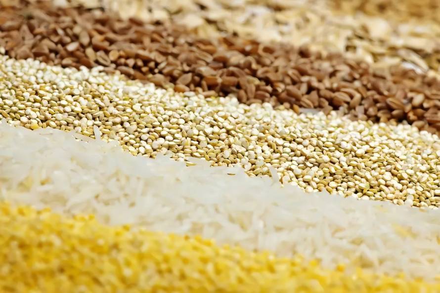
Among the many sources, cereal β-glucan has become the focus of research due to its abundant, safe and reliable source and excellent physicochemical properties. Cereal β-glucan has a variety of physiological functions and effects. It can regulate blood glucose levels and prevent type 2 diabetes [2-4]; lower serum cholesterol levels and prevent cardiovascular disease [5-6]; balance intestinal flora and prevent colon cancer [7-9]; regulate blood pressure [10] and enhance the activity of immune cells [11]. In addition, cereal β-glucan can also be used as an additive in the production of dairy products, ice cream and other foods to improve the sensory quality of these products [12-13].
Currently, the detection method for cereal β-glucan mainly relies on enzymatic methods [14]. In addition, researchers have also developed fluorescence methods [15] and viscosity methods [16] based on the characteristics of β-glucan. Different detection methods differ in terms of cost, accuracy of the test results and testing efficiency, and are therefore suitable for different cereal products or testing requirements.
As an important dietary fiber, the detection of β-glucan is of great importance in the breeding, processing and product development of cereals, especially barley and oats [17-18]. The development of an economical, rapid, accurate, and high-throughput detection method has become a pressing issue for the oat and barley industries. This paper reviews the current principles, methods, and standards for the detection of β-glucan in cereals, with the aim of providing a reference for accurately detecting the β-glucan content of cereals and developing methods to meet specific detection needs.
1 Structure of cereal β-glucan
Grain β-glucan is widely available, high in content, and has a high molecular weight. It is an excellent water-soluble dietary fiber [19]. As a component of plant cell walls, β-glucan can be combined with fluorescent dyes to produce a color, so its distribution in cereal grains can be observed using fluorescent staining techniques [20–22]. Oats and barley are the most common sources of cereal β-glucans, while wheat and rye also contain some β-glucans[23] . Unlike bacterial and fungal β-glucans, cereal β-glucans are linear polysaccharides with a mixture of β-1,3 and β-1,4 linkages. The β-1,4 bonds connect D-glucose monomers to form a cellobiose unit, while the β-1,3 bonds connect these cellobiose units to form β-glucan. The presence of β-1,3 bonds can effectively prevent the molecules from stacking closely and give them a certain degree of water solubility. Therefore, factors such as the ratio of β-1,3 to β-1,4 bonds and the ratio of cellotriose to cellotetraose will affect the physical and chemical properties of β-glucan polysaccharide's physical and chemical properties [23-24].
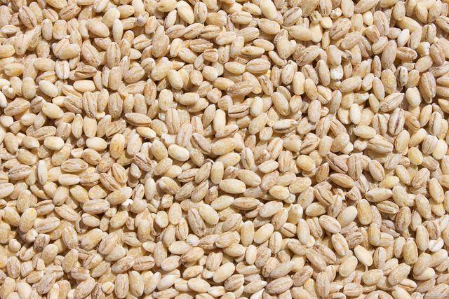
The content and structure of β-glucan in cereals can vary depending on the genetic type and environmental conditions (Table 1). Barley and oats have high β-glucan content, accounting for 2.2% to 8.8% and 1.73% to 5.70% of the dry weight of the grain, respectively, while wheat and rye have β-glucan content of 0.38% to 0.64% and 1.4% to 2.6%, respectively [25-26 ]. In addition, there are certain differences in the ratio of intra-molecular fiber trisaccharides and fiber tetrasaccharides, the ratio of β-1,3 bonds to β-1,4 bonds, and molecular weight of β-glucans in grains. For example, the molecular weight of oat β-glucan is higher, at 180–850 kDa [27], while the molecular weight of rye β-glucan is only 21 kDa [28].
These differences are mainly due to the different genotypes and growth environments of the grains. The structural differences in β-glucan can give it different physical and chemical properties. For example, the ratio of β-1,3 and β-1,4 bonds is related to water solubility, while the molecular weight of β-glucan is related to the viscosity of its solution. A β-glucan solution has a high viscosity and is a non-Newtonian fluid. The higher the concentration and molecular weight of β-glucan, the higher the viscosity of the solution [29]. Beta-glucan can also form gels. The factors that control its gelation behavior are currently unknown, but it is generally believed that the gelation rate is related to the molecular weight of beta-glucan. In addition, beta-glucan can also absorb water and swell. The hydroxyl groups on beta-glucan can form hydrogen bonds with water molecules. At the same time, the internal fiber trisaccharides or fiber tetrasaccharides can be linked by β-1,3 bonds to form binding sites, thereby effectively binding water to them [30].
2. Principle of testing β-glucan in cereals
Currently, there are various methods for testing the β-glucan content of cereals. According to the properties of β-glucan, the principles of these methods can be summarized in four categories: hydrolyzing β-glucan into glucose monomers; specific binding of β-glucan to a dye; using β-glucan's own physical properties; and others.
2.1 Hydrolyzed into glucose monomers
As mentioned above, oat β-glucan is a linear polysaccharide composed of β-1,3 and β-1,4 linkages of D-glucose. It is not easy to directly quantify β-glucan. However, it is more feasible to quantify β-glucan by hydrolyzing β-glucan into D-glucose monomers and then measuring the content of glucose monomers. Generally, biological and chemical hydrolysis methods can be used to hydrolyze β-glucan. The biological hydrolysis method involves the use of specific and highly specific enzymes to cleave the β-1,3 and β-1,4 bonds, thereby degrading the β-glucan into glucose monomers [14]. The chemical method involves the use of a concentrated acid solution at a specific temperature to cleave the β-glucan into glucose monomers [33-34]. Glucose monomers can be converted into colored substances using the oxidase method, and the absorbance of the substance at a wavelength of 510 nm is directly proportional to the glucose content. In addition, the content of glucose monomers can be detected using high-performance liquid chromatography (HPLC) technology. Finally, the β-glucan content can be quantitatively determined by the glucose content.
2.2 Binding with dyes
Beta-glucan can bind to specific substances, changing the color or fluorescence intensity of the complex. This is another main principle for quantitative detection of cereal beta-glucan. For example, if β-glucan binds to the dye Congo red, the binding mechanism may be due to hydrophobic interaction, and the absorbance of the binding solution changes. The amount of β-glucan can be determined by measuring the absorbance change at a wavelength of 550 nm [35]. In addition, β-glucan can also bind to the fluorescent whitening agent Calcofluor, and the amount of β-glucan can be quantitatively detected by measuring the change in the fluorescence intensity of the complex [36]. The specific binding mechanism is currently unclear.
2.3 Physical properties of β-glucan
β-glucan can increase the viscosity of a solution, and it also has strong water absorption properties. These physical properties can be used to detect the β-glucan content in cereals. For a given molecular weight of β-glucan, the viscosity of the solution is directly proportional to the concentration, so the β-glucan content can be detected by measuring the viscosity of the solution [16]. It is worth noting that the molecular weight of β-glucan also affects the viscosity of the solution. The higher the molecular weight, the higher the viscosity. Therefore, when using this principle for detection, attention must also be paid to the effect of molecular weight. The strong water absorption of β-glucan can increase the water retention of the cereal system, that is, the higher the β-glucan content, the greater the water retention of the system. Based on this principle, Niu et al. [37] developed a new method for the detection of oat β-glucan using solvent retention capacity.
2.4 Other
In addition to the above principles, there are also spectroscopic methods and enzyme-linked immunosorbent assays for the detection of β-glucan. Spectroscopy uses near-infrared spectroscopy, which is based on the fact that certain chemical bonds in β-glucan will produce vibrational absorption under near-infrared light, in accordance with Lambert–Beer's law, i.e. the intensity of the absorption peak is proportional to the concentration of the substance to be measured [38]. The principle of enzyme-linked immunosorbent assay is to develop specific monoclonal antibodies that specifically bind to β-glucan as a substrate, and quantitatively detect the β-glucan content through a colorimetric reaction [39].
3. Methods for the detection of β-glucan in cereals and related standards
β-glucan is widely used in foods and other fields, and has important physiological activities. Therefore, the β-glucan content needs to be detected in all aspects of the oat industry (breeding, processing, product development, etc.). Table 2 summarizes the principles, advantages and disadvantages, and relevant standards of the current β-glucan detection methods.
3.1 Enzyme method
At present, the enzyme method is the most commonly used method for detecting β-glucan and has been developed into a kit that has become a classic commercial detection method. This method was first proposed and improved by McCleary in 1985 [14] and can be used to detect the content of β-glucan in cereals and their products. The enzymatic method is an internationally recognized method for the detection of β-glucan and has been certified by many international authorities such as the American Association of Analytical Chemists (AOAC International, 995.16) [40] and the American Association of Cereal Chemists (AACC, 32-23.01) [41].
This method uses lichenase and β-glucosidase to hydrolyze (Figure 1). First, the β-glucan in the grains or their products is dissolved in boiling water, and then specifically hydrolyzed into small molecular oligosaccharides by lichenase (lichenase). After that, β-glucosidase is used to cleave it into individual glucose molecules. By measuring the glucose quantitative detection of β-glucan, even if glucose is converted to a colored substance with glucose oxidase buffer, and the amount is quantitatively detected at a wavelength of 510 nm. This method is highly specific and not easily interfered with by other polysaccharides. The detection results are highly accurate and stable, and it is the most widely used method for detecting β-glucan. However, high-purity and highly specific licheninase and glucosidase are expensive, and the cost of detection is high.
In order to reduce the amount of sample used, save reagent consumption and reduce testing costs, Hu and Burton [42] made some improvements to this testing method. They used a 96-well plate method to determine the β-glucan content of 21 different oat samples, reducing the amount of lichenase and β-glucosidase used in the traditional enzymatic method by 25%, reducing the cost of testing each sample by 22% and labor costs by 25%. Afterwards: Motilva et al. [43] used a micro method based on the Megazyme enzymatic assay to further optimize the detection of samples with β-glucan content ranging from 0.27% to 75%. The traditional Megazyme enzyme method requires 0.1 mL of β-glucosidase to be added after the sample is hydrolyzed by lichenase to hydrolyze it into glucose monomers, and the reaction is colored with 3 mL of GOPOD (glucose oxidase buffer) reagent. The improved micro method only requires 20 μL of β-glucosidase and 210 μL of GOPOD. Tests have shown that the results are not significantly different from the enzymatic method.
3.2 Chromatography
Chromatography can be used to determine the β-glucan content of cereals, as the polysaccharides can be broken down into smaller substances under acidic conditions at a certain temperature. The concentration of the acid solution, the temperature and the duration of the hydrolysis affect the efficiency of the hydrolysis, leading to incomplete hydrolysis or damage to the structure of the monosaccharide products. The hydrolysate glucose can be further analyzed by gas chromatography (GC) or high performance liquid chromatography (HPLC). Johansson et al. [33] compared the hydrolysis of β-glucan by three acid solutions (HCl, TFA and H 2 SO) at different acid concentrations, temperatures and hydrolysis times. The results showed that β-glucan was not hydrolyzed under the weakest acidic conditions (37 °C, pH = 1, simulating the acidic conditions of human gastric juice), while the three acid solutions obtained the same glucose content when hydrolyzing β-glucan at 120 °C , for 1 h, to obtain the same glucose content. The results of this method are similar to those of the enzymatic method, with high accuracy. However, it requires the use of expensive chromatographic equipment, and the experimental conditions of the pretreatment process (high temperature, strong acid) pose a high safety risk. These factors limit the use of this method to a certain extent.

3.3 Congo red method
The Congo red dye can form a complex with β-glucan. Congo red is a red dye that absorbs light in the ultraviolet-visible region. After β-glucan is added, the absorbance of the complex at a wavelength of 550 nm changes with the concentration of β-glucan. When using this method to detect the β-glucan content of cereals, a standard β-glucan of different concentrations needs to be added to a certain concentration of Congo red solution, mixed to establish a standard β-glucan concentration curve, and then the β-glucan content in the sample can be measured [35]. The Congo red dye used in this method is relatively inexpensive, so the cost is low. However, the binding of Congo red to β-glucan is not specific, and when it is applied to cereals, it is easily interfered with by other water-soluble polysaccharides such as pentosans, which affects the measurement results. Therefore, this method has certain limitations in accuracy.
3.4 Fluorescence method
The fluorescence method is also one of the commonly used methods for quantifying β-glucan. This method is based on the change in the fluorescence intensity of the solution, that is, the fluorescent agent Calcofluor and β-glucan can specifically form a complex, and the fluorescence intensity of the system solution is related to the concentration of β-glucan. Based on this principle, Jorgensen [44] designed an automatic flow injection analysis system in 1988, which uses wet analytical chemistry to mix the sample solution with the fluorescent agent to detect the β-glucan content in the sample.
This method has been widely used to detect β-glucan in liquid samples such as beer and wort. The method requires the sample containing β-glucan to be diluted into a solution, and a standard β-glucan solution is used to establish a concentration-fluorescence intensity standard curve, from which the β-glucan content in the sample can be obtained. Compared with the traditional enzyme method, the fluorescence method is less expensive. At the same time, the results of the fluorescence method are significantly correlated with the enzyme method, and the accuracy is higher. However, the fluorescent agent Calcofluor is not very stable and is easily decomposed by light. In addition, the composition of the cereal system is complex, and substances such as protein and starch can also interfere with the results. These factors limit its application in the determination of β-glucan in cereals to a certain extent.

3.5 Viscosity method
Beta-glucan powder is highly viscous, and the higher the concentration of the solution, the higher the viscosity. For oat flour and barley flour, the apparent viscosity of the slurry mainly depends on the content of beta-glucan, while starch and protein have only a small effect. Colleoni-Sirghie et al. [16] prepared oat flour homogenates by adding oat flour to deionized water, a silver nitrate solution (to inhibit endogenous β-glucanase) and an alkali solution (to dissolve water-soluble and water-insoluble β-glucans), respectively, and studied the relationship between the apparent viscosity of the homogenate and the β-glucan content. method (PLS) to predict the β-glucan content. The results showed that the apparent viscosity of the oat flour homogenate using silver nitrate solution can be used to predict the β-glucan content very well, and the results are very close to those obtained by the enzymatic method. The viscosity method is simple in principle, low in cost, and easy to operate. However, the viscosity of β-glucan solutions is also affected by molecular weight, and samples with similar molecular weights need to be used when using this method. Therefore, this method can be used to detect the content of β-glucan with the same molecular weight, such as the β-glucan content in different products made from the same oat kernel.
3.6 Solvent retention capacity method
Solvent retention capacity (SRC) refers to the ability of wheat flour to retain a certain amount of solvent under a certain centrifugal force, and can be used to measure the quality characteristics of wheat flour and its products. The principle is that the polymer molecules in wheat flour, such as proteins, starch, pentosans, etc., can swell to varying degrees in different solvents. The SRC test can use four solvents: water, 5% (w/w) aqueous lactic acid solution, 5% (w/w) aqueous sodium carbonate solution, and 50% (w/w) aqueous sucrose solution. These four solvents correspond to different polymer components in wheat flour. The water SRC reflects the swelling capacity of all polymer components in the flour; the lactic acid SRC has a similar pH value during the dough fermentation process and is therefore related to the swelling capacity of glutenin; the sodium carbonate SRC solution has a higher pH value, which can dissociate the hydroxyl groups of starch. Damaged starch can absorb water and expand in this solution, so this SRC is related to the content of damaged starch . The sucrose SRC solution is concentrated and neutral, and this solution amplifies the swelling effect of the arabinoxylan network, so it is related to the content of wheat pentosans [45].
Beta-glucan is a large molecular polymer with many hydroxyl groups and strong water absorption. The beta-1,3-linked fructan units can bind water molecules, so it has strong water absorption and swelling capacity. Niu et al. [37] first applied the wheat SRC method to oats, studied the relationship between different solvents and the swelling characteristics of oats, and selected a calcium chloride solvent from this to reflect the β-glucan content of oats. This method can directly add the corresponding solvent (25 g) to 5 g of oat flour when testing for β-glucan content, and after centrifugation, the β-glucan content can be predicted by measuring the weight of the solvent retained by the oat flour. The principle is simple and the operation is easy. However, other macromolecular polymers in the cereal system can also cause swelling, and when the β-glucan content is low, it is not easy to detect. Therefore, the accuracy of this method needs to be further optimized. This method can be used to predict the approximate range of oat β-glucan content, and therefore can be used for preliminary screening of β-glucan content in breeding and other processes. In addition, our previous research results also show that there is also a significant correlation between oat flour SRC and β-glucan molecular weight [46], which provides some ideas for rapid detection of β-glucan molecular weight.
3.7 Near-infrared method
NIR spectroscopy has been widely used in agriculture, pharmaceuticals, polymer manufacturing and food quality testing. In the NIR spectral region, chemical bonds in molecules (such as C-H, N-H, O-H bonds) have characteristic peaks at specific wavelengths. The composition of a substance can be detected by analyzing the frequency doubling peaks of one or more molecules in a substance's spectral region. When using near-infrared methods to predict the content of a component, the near-infrared spectrum of a sample set must first be collected and the original spectrum pre-processed. Mathematical analysis is then used to establish a prediction model for the component, and finally the model is tested and converted for application [47]. At present, some domestic and foreign studies have reported the use of near-infrared methods to detect β-glucan in cereals. The samples used in these studies were mainly crushed cereal flour.
Some studies used intact grain kernels for testing, and these studies used the results of the Megazyme enzymatic assay as a reference value for mathematical modeling [48-54]. Schmidt et al. [38] evaluated the potential of near infrared spectroscopy for the determination of β-glucan in barley. They analyzed 107 barley samples (whole grain and barley flour, respectively) using four different near infrared instruments. First, the spectra were collected and the raw spectra were preprocessed. A partial least squares (PLS) method was used to establish a near infrared prediction model for β-glucan in barley. The results show that all tests show a certain applicability for β-glucan monitoring (R2 > 0.78). This method is fast and simple, and can be used to test large batches of samples in a short period of time. It should be noted that the accuracy of the NIR method is affected by many factors, including the accuracy of the chemical reference value of the sample, the content of the index to be measured in the sample and whether it is normally distributed, high background and overlapping peaks, sample size, and the mathematical model selected.
The β-glucan structure is simple, and the chemical bonds within the molecule can easily overlap with other components of oats (starch, protein, etc.), affecting accuracy. This method is suitable for testing when the sample size is large, but it still requires a large number of samples with known chemical reference values (the sample size generally needs to be greater than 100) to establish an NIR quantitative model.
3.8 Enzyme-linked immunosorbent assay
The enzyme-linked immunosorbent assay (ELISA) is a common biochemical analysis method. This method uses specific antibodies to detect the presence of ligands in liquid samples. ELISA can also be used to determine the trace levels of β-glucan in cereals. Rampitsch et al. [39] developed different monoclonal antibodies and studied their degree and specificity of reaction with β-glucans from oats and barley. The results of the ELISA for the detection of β-glucan from commercial oats were linear within a certain range (1–20 ng β-glucan·mL-1). In addition, they optimized the experimental conditions for the ELISA and developed a method that can save time and labor costs for the detection of large quantities of samples. In theory, the ELISA method can detect β-glucan at the nanogram level and can be used for trace testing when the sample size is small. However, due to the extremely high specificity and sensitivity of this method, it may not be able to identify all β-glucans with similar structures, and its applicability needs to be further studied.
4 Conclusion
Grain β-glucan is an important source of dietary fiber, and the β-glucan content needs to be detected in many processes such as breeding and processing of grains, especially oats and barley. Among the currently developed methods for detecting β-glucan in grains, enzymatic methods, chromatographic methods, and fluorescence methods can be used to accurately quantify β-glucan content. The viscosity method and the solvent retention capacity method are simple to operate and can be used for preliminary screening of β-glucan content. Near-infrared spectroscopy is fast and can be used for batch testing when there is a large sample size. One can reasonably choose a testing method that meets their needs by considering factors such as accuracy, operational efficiency, and cost.
Reference:
[1] AHMAD A , ANJUM F M , ZAHOOR T , et al.Beta glucan : a valuable functional ingredient in foods [J] . CriticalReviews in Food Science and Nutrition , 2012 , 52(3) :201.
[2] REGAND A , CHOWDHURY Z , TOSH S M , et al.The mo- lecular weight , solubility and viscosity of oat beta-glucan affect human glycemic response b y modifying starch digestibility [J] . FoodChemistry , 2011 , 129(2) :297.
[3]SHEN R L , CAI F L , DONG J L , et al.Hypoglycemic effects and biochemical mechanisms of oat p roducts on streptozoto- cin-induced diabetic mice [J] . Journal of Agricultural and FoodChemistry , 2011 , 59(16) :8895.
[4] ZHAO F , LIU H P , LIUXQ , et al.Effectof oat branβ-glucan on hypoglycemic and antioxidant [J] . Journalof Food Safety andQuality , 2015 , 6(6) :2131.
[5] WOLEVER T M S , GIBBS A L , BRAND-MILLER J , et al. Bioactive oat β-glucan reduces LDL cholesterol in caucasians and non-caucasians [J] . NutritionJournal , 2011 , 10(1) : 1.
[6] NING H Z , WANG S B , LIU Y L , et al.Effect of oat glucan on vascular endothelial active substances and inflammatory re- sponse markers in hyp ercholesterolemic rats [J] . Chinese Journalof Food Hygiene , 2011 , 23(3) :233.
[7] CHOROMANSKA A , KULBACKA J , HARASYM J , et al. Anticancer activity of oat β-glucan in combination with elec- troporation on human cancer cells [J] . Acta Poloniae Phar- maceutica-Drug Research , 2017 , 74 : 616.
[8] MRTENSSON O , IRASTORZA A , HOLST O , et al.Effects of fermented , ropy , non-dairy , oat-based p roducts on serum lipids and the faecal excretion of cholesterol and short chain fatty acids in germfree and conventional rats [J] . Nutrition Research , 2002 , 22(12) : 1461.
[9] ZHANG L , CHEN D W , YU B , et al.Short-term adding high- level oat β-glucan , microcrystalline cellulose and their mixture in diets affects growth p erformance , organ indexes and fecal bacterial community structure of mice [J] .ChineseJournalof Animal Nutrition , 2017 , 29(7) :2407.
[10]JAYACHANDRAN M , CHEN J , CHUNG S S M , et al. A critical review on the im pacts ofβ-glucans on gut microbiota and human health [J] . The Journal of Nutritional Bio- chemistry , 2018 , 61 : 101.
[11]CHANGCF , CHAN W K , SZED M Y.Theeffectsofβ-glu- can on human immune and cancer cells [J] . Journal of He - matology & Oncology, 2009 , 2(1) : 1.
[12] WANG F M , FAN M S , ZHENG K K.Nutritive value of oat β-glucan and the factors affecting its accumulation [J] . Jour- nalofTriticeaeCrops , 2005 , 25(2) : 116.
[13]ZHU F , DU B , XU B.A critical review on p roduction and in- dustrial applications of beta-glucans [J] . Food Hydrocol- loids , 2016 , 52 :275.
[14] MCCLEAR B V , GLENNIE-HOLMES M.Enzymic quantifi- cation of(1→ 3)(1→ 4) -β-D-glucan in barley and malt [J] . Journalof the Instituteof Brewing , 1985 , 91(5) :285.
[15] RIEDER A , KNUTSEN S H , BALLANCE S , et al.Cereal β- glucan quantification with calcofluor-application to cell cul- ture supernatants [J] . Carbohydrate Polymers , 2012 , 90 (4) : 1564.
[16]COLLEONI-SIRGHIE M , JANNINK J L , KOVALENKO I V , et al.Predictionofβ-glucan concentration based on viscos- ity evaluations of raw oat flours from high β-glucan and tra-ditional oat lines [J] .CerealChemistry , 2004 , 81(4) :434.
[17] HU X Z , WEI Y M , REN C Z. Oat quality and p rocessing [ M] .Beijing:Science Press , 2009 : 10-15.
[18]REN C Z , HU X Z , China ’s oat and buckwheat industry “ Twelfth Five-Year”development report (2011-2015) [ M] . Xi’an :Shaanxi Science and Technology Press , 2016 :32-48.
[19] DU B , MEENU M , LIU H , et al. A concise review on the molecular structure and function relationship of β-glucan [J] . International Journal of Molecular Sciences , 2019 , 20 (16) :4032.
[20] HU X , ZHAO J , ZHAO Q , et al.Structure and characteristic of β-glucan in cereal:A review [J] . Journalof Food Process- ing and Preservation , 2015 , 39(6) :3145.
[21] WOOD P J . Oat β-glucan : structure , location and properties [J] .Chemistry andTechnology , 1986 : 121.
[22]CUI W , WOOD P J , BLACKWELL B , et al.Physicochemical properties and structural characterization b y two-dimensional NMR spectroscopy of wheat β-D-glucan-comparison with other cereal β-D-glucans [J] .Carbohydrate Polymers , 2000 , 41(3) :249.
[23]LAZARIDOU A , BILIADERIS C G.Molecular aspects of ce- real β-glucan functionality:Physical properties , technological applications and physiological effects [J] . Journal of Cereal Science , 2007 , 46(2) : 101.
[24] WOOD P J , WEISZ J , BEER M U , et al.Structure of(1-3)(1- 4) -β-D-glucan in waxy and nonwaxy barley [J] . Cereal Chemistry , 2003 , 80(3) :329.
[25]IZYDORCZYK M S , BILIADERIS C G.Structural and func- tional aspects of cereal arabinoxylans and β-glucans [ M]// Developments in Food Science.Elsevier , 2000 , 41 :361-384.
[26] HAVRLENTOVA M , KRAIC J A N.Content of beta-d-glu- can in cereal grains [J] . Journalof Food and Nutrition Re- search(SlovakRepublic) ,2006 , 45(3) : 97.
[27]SKENDI A , BILIADERIS C G , LAZARIDOU A , et al.Struc- ture and rheological properties of water soluble β-glucans from oat cultivars of Avena sativa andAvenabysantina [J] . Journalof Cereal Science , 2003 , 38(1) : 15.
[28]BHM N , KULICKE W M.Rheological studies of barley(1 → 3)(1→ 4) -β-glucan in concentrated solution : Mechanistic and kinetic investigation of the gel formation [J] . Carbohy - drate Research , 1999 , 315(3-4) :302.
[29] MORRIS E R. Polysaccharide solution properties : origin , rheological characterization and im plications for food sys- tems [J] . Frontiers in Carbohydrate Research 1 , Food Ap- plications , 1989 : 132.
[30]ZIELKE C , STRADNER A , NILSSON L.Characterization of cereal β-glucan extracts :Conformation and structural aspects [J] . Food Hydrocolloids , 2018 , 79 :218.
[31] LI W , CUI S W , KAKUDA Y. Extraction , fractionation , structural and physical characterizationofwheatβ-D-glucans [J] .Carbohydrate Polymers , 2006 , 63(3) :408.
[32] VAIKOUSI H , BILIADERIS C G , IZYDORCZYK M S.Solu- tion flow behavior and gelling properties of water-soluble barley(1→ 3 , 1 → 4) -β-glucans varying in molecular size [J] . Journalof Cereal Science , 2004 , 39(1) : 119.
[33]JOHANSSON L , VIRKKI L , ANTTILA H , et al.Hydrolysis of β-glucan [J] . FoodChemistry , 2006 , 97(1) : 71.
[34] YU Y J , DAI J , ZHU S , et al. Determination of β-D-glucan inlentinan b y acidic and enzymatic hydrolysis coupled HPLC [J] . Food and Fermentation Industries , 2012 , 38(7) : 148.
[35] ZHANG J , DU X F , RAO Y Q. Measurement of beta-glucan form oats b y congo red [J] . Journal ofAnhuiAgricultural University , 2007 , 34(1) :23.
[36] WU J , DENG X , TIAN B , et al.Interactions between oat β- glucan and calcofluor characterized b y spectroscopic method [J] . JournalofAgricultural and Food Chemistry , 2008 , 56 (3) : 1131.
[37] NIU Q , PU Y , LI X , et al.Solvent retention capacities of oat flour [J] . International Journal of Molecular Sciences , 2017 , 18(3) :590.
[38] SCHMIDT J , GERGELY S , SCHNLECHNER R , et al. Comparison of different types of NIR instruments in ability to measure β-glucan content in naked barley [J] . Cereal Chemistry , 2009 , 86(4) :398.
[39] RAMPITSCH C , AMES N , STORSLEY J , et al. Develop- ment of a monoclonal antibody-based enzyme-linked immunosorbent assay to quantify soluble β-glucans in oats and barley [J] . Journal of Agricultural and Food Chemistry , 2003 , 51(20) :5882.
[40] AOACInternational.AOACOfficialMethod995.16:β-D-glu- can in barely and oats [S] . AOAC International , Gaithers- burg, Md , 2000.
[41] AACC International.Approved methods of analysis , 10th Ed. Method 32-20.01 [S] .AACCI:St.Paul , MN , 2005.
[42] HU G , BURTON C.Modification of standard enzymatic pro- tocol to a cost ‐ efficient format for mixed linkage(1→ 3 , 1 → 4) -β-d-glucan measurement [J] .CerealChemistry , 2008 , 85(5) : 648.
[43] MOTILVA M J , SERRA A , BORRAS X , et al.Adaptation of the standard enzymatic p rotocol( Megazyme method) to mi- croplaque format for β-( 1 , 3) (1 , 4) -d-glucan determination in cerealbased sampleswithawide rangeofβ-glucan content [J] . Journalof Cereal Science , 2014 , 59(2) :224.
[44]JORGENSEN K G , AASTRUP S.Quantification of high mo- lecular weight( 1 → 3) ( 1 → 4) -β-D-glucan using calcofluor complex formation and flow injection analysis.II.Determina- tion of total β-glucan content of barley and malt [J] . Carls- berg Research Communications , 1988 , 53(5) :287.
[45] KWEON M , SLADE L , LEVINE H.Solvent retention capacity(SRC) testing of wheat flour:Principles and value in predicting flour functionality in different wheat-based food processes and in wheat breeding:A review [J] .CerealChem- istry , 2011 , 88(6) :537.
[46]ZHANG K , LI X , MA Z , et al.Solvent retention capacity of oat flour:Relationship with oat β-glucan content and molecu- lar weight [J] . Food Hydrocolloids , 2019 , 93 : 19.
[47] GAO R Q , FAN S F.Principles and applications of modern near in frared spectroscopic techniques [J] . PrecInstOpto- EelectEngineering , 2002(3) : 11.
[48]SOHN M , HIMMELSBACH D S , BARTON F E , et al.Near infrared analysis of whole kernel barley:Comparison of three spectrometers [J] .Applied Spectroscopy , 2008 , 62(4) :427. [49] SCHMIDT J , GERGELY S , SCHONLECHNER R , et al.
Comparison of different types of NIR instruments in ability to measure β-glucan content in naked barley [J] . Cereal Chemistry , 2009 , 86(4) :398.
[50]BELLATO S , FRATE V D , REDAELLI R , et al.Use of near infrared reflectance and transmittance coupled to robust cali- bration for the evaluation of nutritional value in naked oats [J] . JournalofAgricultural and Food Chemistry , 2011 , 59 (9) :4349.
[51]LIU H , ZHOU H , REN G.Using Fourier transform near in- frared spectroscopy to estimate the nutritional value in whole and milled naked oats [J] . Journal of Near Infrared Spec- troscopy , 2014 , 22(2) : 93.
[52] GRACIA M B , ARMSTRONG P R , RONGKUI H , et al. Quantification of beta-glucans , lipid and p rotein contents in whole oat grains(Avena sativa L. ) using near infrared re- flectance spectroscopy [J] . Journal of Near Infrared Spec- troscopy , 2017 , 25(3) : 172.
[53] RINGSTED T , RAMSAY J , JESPERSEN B M , et al.Long wavelength near-infrared transmission spectroscopy ofbarley seeds using a supercontinuum laser:Prediction of mixed-link- age beta-glucan content [J] . Analytica Chimicaacta , 2017 , 986 : 101.
[54] KRISHNAN P , CAFFE-TERML M , PAUDEL D. A single analytical platform for the rapid and simultaneous measure- ment of p rotein , oil and beta glucan contents of oats using near infrared reflectance spectroscopy [J] . Cereal Foods World , 2018 , 17.
[55] ICC. ICC Standard methods of the international association for cereal chemistry, 166 [S] .The Association , Vienna , 1996.
[56] European Brewery Convention. Analytica-EBC , 5th ed [S] . Fachverlag Hans Carl:Nurnberg, Germany, 2007.
[57] RACI.Official testing methods of the cereal chemistry divi- sion [S] . Royal Australian Chemical Institute , Melbourne , Australia.
[58] Codex Committee on Methods of Analysis and Sampling.CRD16from the thirty-third session of the Codex Committee on Methods of Analysis and Sampling [S] . FAO : Rome , 2012.
[59] Agricultural Products Processing Bureau , Ministry of Agri- culture of the People's Republic of China.Industry Standards of the Ministry of Agriculture of the People's Republic of China:NY/T 2006-2011Determinationofβ-glucan content in cereals and their p roducts [S] .Beijing:China Standard Press , 2011.


