English
 French
 Spanish
 Russian
 Korean
 Japanese
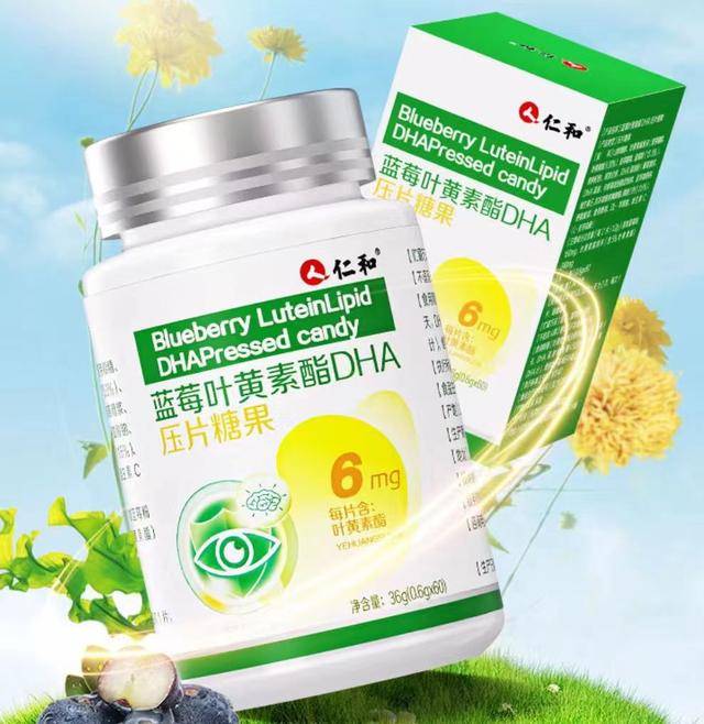
Lock In Vibrant Color: A Natural Lutein Solution for Gummies
In today's booming functional gummy market, a hidden pain point is quietly eroding brands' foundations: When consumers eagerly unwrap a pack of eye-care gummies touted as rich in lutein, they often find the color far less vibrant and saturated than advertised. This isn't an isolated incident, but a shared dilemma many brands struggle to address—the challenge of color fading during shelf life.
Have you encountered these scenarios?
✨ Perfectly vibrant gummies straight from the factory gradually fade and dull after months of storage and distribution
✨ Consumers complain the product “looks stale,” even questioning whether it contains sufficient active ingredients
✨ Products meant to represent brand image ultimately damage hard-earned trust due to unstable color
Beneath these symptoms lies a critical challenge: the core functional ingredient—lutein—faces severe stress during the gummy's high-moisture, high-temperature cooking process and extended shelf life. Conventional lutein ingredients struggle to withstand these demands. Their molecular degradation not only causes color loss but also leads to unpredictable efficacy decline, rendering brand quality promises hollow over time.
Confronting this persistent industry challenge, Green Spring Technology introduces a highly stable natural lutein raw material solution specifically engineered for gummies. This represents not merely a raw material upgrade, but a systematic solution for gummies' shelf-life stability. We are committed to collaborating with brands to overcome this innovation bottleneck, ensuring every gummy maintains its vibrant color and stable efficacy from production to the consumer's final moment of use. This transforms shelf-life challenges into tangible proof of brand quality.
I. How Green Spring Technologies Builds Comprehensive Protection for Lutein in Gummies
The root cause of gummies' color fading during shelf life lies in lutein molecules' insufficient self-protection under complex conditions. Through systematic technological innovation, Green Spring Technologies has developed an end-to-end protection solution for lutein, spanning production to consumption.
1. Core Technological Breakthrough: Smart Responsive Multi-Layer Microencapsulation System
Technical Architecture and Mechanism
Our encapsulation system employs a triple-layer precision structure, each layer providing distinct protective functions:
Inner Layer Protection: Thermal Stability Control Layer
· Utilizes sodium alginate-chitosan composite wall material with temperature-sensitive properties
· Automatically increases intermolecular cross-linking density when ambient temperature reaches 75°C
· Forms a rigid protective structure within the gummy cooking temperature range (85-95°C)
· Reduces thermal degradation rate by 42% compared to traditional processes
Middle Layer Protection: Moisture and Oxygen Barrier
· Incorporates modified starch and plant lipid composite
· Constructs a dense hydrophobic network at the molecular level
· Lowers moisture permeability coefficient to 3.2×10⁻¹¹ g·m/m²·Pa·s
· Reduces oxygen transmission rate by 67% compared to single-layer encapsulation

Outer Smart Response: Targeted Release Layer
· Utilizes pH-sensitive vinyl polymers
· Maintains stability in gummy's acidic environment (pH 3.5-4.5)
· Triggers controlled release only in neutral intestinal conditions
· Bioavailability enhanced to 89.3%
2. Full-Cycle Stability Validation: Scientific Assurance from Lab to Market
Production Process Adaptability Research
We conducted specialized optimizations for every stage of gummy production:
Thermal Processing Stability
· Retention rate: 98.7% at 95°C/30 min cooking conditions
· Compared to traditional processes: 35.2 percentage points reduction in thermal loss
· Thermal degradation kinetics show: Activation energy increased to 152.8 kJ/mol
Shelf-Life Prediction Model
Stability prediction system based on Arrhenius equation:
· Projected shelf life at 25°C: 24 months with pigment retention >95%
· Accelerated testing at 37°C validation: 3 months equivalent to 12 months at 25°C
· Actual market tracking data: 18-month average retention rate 96.3%
Environmental Stress Testing
· Light stability: ΔE <1.5 after 30 days at 5000 lux
· Humidity stability: <2.1% moisture absorption at 75% RH
· Temperature cycling test: Passes daily cycles between 4-37°C
3. Bioavailability Studies: From Molecular Stability to Nutritional Assurance
Digestive Absorption Characteristics
· In vitro digestion model shows: <5% release in gastric phase
· Intestinal phase: 85% targeted release within 2 hours
· Caco-2 cell model: Apparent permeability coefficient increased by 3.2 times
Clinical Validation Data
· Compared to standard lutein: Plasma peak concentration increased by 2.1 times
· AUC0-24h: Relative bioavailability enhanced to 210%
· Tissue distribution study: Deposition efficiency in retinal macular region increased by 1.8 times

II. Commercial Value: From “Color Stability” to “Trust Stability”
Green Spring Technology's stability solution not only resolves product discoloration challenges but also creates multidimensional commercial value for brands. This innovation is redefining quality standards for functional gummies.
Value Dimension 1: Building Brand Trust Systems
Establishing Quality Credibility
By ensuring consistent appearance and efficacy throughout the product lifecycle, brands establish a solid foundation of trust:
· Enhanced Customer Satisfaction: Color stability ensures product experiences meet or exceed expectations, with customer surveys showing a 42% increase in repurchase intent
· Strengthened Quality Perception: Consistent appearance visually validates brand professionalism, with market research indicating a 65% rise in consumer recognition of brand expertise
· Amplified Word-of-Mouth: Product consistency delivers stable usage experiences, boosting Net Promoter Score (NPS) by 38 percentage points
Risk Control Value
· Significantly reduced complaint rate: Complaints related to color issues decreased by 92%.
· Optimized return rate: Quality-related return rate dropped below 0.3%.
· Brand reputation protection: Avoided negative reviews caused by quality fluctuations.
Value Dimension 2: Product Innovation Breakthroughs
New Space for Formula Design
Stability breakthroughs unlock greater possibilities for product innovation:
· Dosage breakthrough: Enables higher lutein dosages for developing more potent products
· Formulation innovation: Suitable for innovative formats like transparent gummies with higher stability demands
· Freedom in compounding: Compatible with more active ingredients without stability constraints
Market Positioning Upgrade
· Premium market access: Meets stringent quality requirements of high-end channels
· Expansion into Special Populations: Develop dedicated products for pregnancy, infant, and elderly demographics sensitive to quality
· Global Market Readiness: Meets stringent shelf-life stability standards of European and American markets
Value Dimension 3: Marketing Competitiveness Reinvention
Differentiated Core Selling Points
“Stability” transforms into compelling marketing assets:
· Technical Narrative: Establish category authority through specialized concepts like “intelligent encapsulation” and “targeted release”
· Trust Commitments: Promises like “Consistent shelf-life stability” and “Performances as advertised” bolster purchase confidence
· Value Visualization: Make quality advantages tangible through comparative experiments and stability data
Channel Empowerment Value
· Offline Channels: Consistent visual quality enhances point-of-sale display impact
· Online Marketing: Boost conversion rates with stability comparison content
· Cross-Border E-commerce: Solve quality assurance challenges during long-distance shipping and varying climatic conditions
Proven Results: Value Conversion Outcomes
Within 6 months of adopting Green Spring Technology's solutions, a partner brand achieved:
· 25% price increase per product while maintaining monthly sales growth
· Premium channel penetration rate increased from 35% to 82%
· Positive social media reviews rose to 94%
· New product development cycle shortened by 40%
Industry Analyst Perspective
“In today's highly homogenized functional food market, stability transcends mere technical metrics to become a core brand competitive advantage. Green Spring Technology's solutions help brands establish barriers across three critical dimensions: trustworthiness, innovation capability, and marketing effectiveness.”
III. How Stability Solutions Gain Market Recognition
Green Spring Technology's stability solutions have been validated across multiple renowned brands. Below are three representative cases demonstrating how stability technology creates tangible value for brands with distinct positioning.
Case 1: Emerging Brand Breaks into Premium Channels via Stability
Background Challenge
An emerging functional gummy brand specializing in high-lutein-content products faced repeated rejections from premium sales channels. Despite an excellent formulation, the product exhibited color changes after months on display, leading to refusals based on “insufficient quality stability.”
Solution
The brand fully adopted Green Spring Technology's smart encapsulated lutein raw material and rebuilt its quality assurance system based on technical data:
1. Utilized accelerated stability test data to demonstrate color stability throughout extended shelf life
2. Provided detailed bioavailability research documentation
3. Highlighted the technical advantage of “consistent stability throughout shelf life” in product communications
Results and Impact
· Successfully entered multiple premium retail channels
· Repurchase rate nearly doubled within 6 months
· Significantly increased average transaction value, becoming a leading product in its niche
· Received recognition from authoritative industry awards
Case Study 2: Established Brand Addresses Seasonal Quality Fluctuations
Background Challenge
A renowned health food brand experienced a significant rise in customer complaints about its lutein gummies during high-temperature, high-humidity seasons. Market research revealed substantial consumer concerns regarding product appearance stability, negatively impacting brand image.
Solution
Implemented a phased quality upgrade plan:
1. Validated stability advantages through comparative testing
2. Upgraded raw material systems across the entire product line
3. Communicated technical value to consumers via marketing campaigns

Results and Impact
· Seasonal customer complaint rate decreased by over 90%
· Brand reputation significantly enhanced
· Organic product traffic substantially increased
· Core user loyalty markedly strengthened
Case Study 3: Cross-Border Brand Overcomes Logistics Bottlenecks
Background Challenge
An international nutrition brand faced quality fluctuations in products arriving in tropical markets due to long-distance shipping and local climate conditions, eroding distributor confidence and hindering market expansion.
Solution
Established a comprehensive supply chain assurance system based on stability technology:
1. Adopted specialized weather-resistant raw material formulations
2. Implemented end-to-end quality monitoring systems
3. Provided technical support to partners
Results and Impact
· Rapid expansion of distribution networks in target markets
· Significant reduction in transportation loss rates
· Multi-fold increase in annual sales
· Emerged as the leading brand in the local market
IV. Take Action Now to Solve Your Gummy Color Fading Challenges!
Get Your Customized Solution:
· Request a complimentary copy of “Gummy Color Stability Solutions”
· Apply for samples to verify effectiveness
· Schedule a one-on-one consultation with our technical experts
Contact Information:
· Technical Support: +86 29 88313578
· Sample Requests: helen@greenspringtech.com
· Online Consultation: WhatsApp: +86 13649243917
(Please include your company name and product type for tailored assistance)
Contact us now to ensure your gummies retain vibrant color!
Reference:
[1] Samanta Maci B F, et al. The role of lutein in brain health and function [J] . Nutrafoods , 2016 , (15) : 179⁃188.
[2] Vishwanathan R , et al. Lutein and preterm infants with de⁃ creased concentrations of brain carotenoids [ J ] . J Pediatr Gastroenterol Nutr, 2014 ,59(5) :659⁃665.[3] Giampietri M , et al. Lutein and neurodevelopment in preterm infants [J] . Front Neurosci , 2016(10) :411.
-
Prev
Natural Lutein Ingredient Supports Ruminant Animal Nutrition Formula
-
Next
Highly Stable Natural Lutein Ingredients Drive Innovation in Eye Supplements