

 English
 French
 Spanish
 Russian
 Korean
 Japanese
Study on Marigold Extract Lutein Good for Eyes
Many compounds with antioxidant structures found in food or vegetables have pharmacological effects such as anti-inflammatory, regulating blood lipids, and inhibiting angiotensin II, such as polyphenols, flavonoids, isoflavones, allicin, lycopene, phytosterols, etc. [1]. Among them, the role played by lutein in the prevention of cardiovascular disease has gradually received people's attention [2]. At the same time, as the main pigment that constitutes the macula of the retina, its protective effect on the retina has been confirmed, and therefore its position in eye health care is becoming increasingly prominent. This paper reviews the research progress of lutein's eye health care and adjuvant therapeutic effects.
1 Source and properties of lutein
Lutein is widely found in green vegetables (such as kale, spinach, broccoli, peas, lettuce, etc.) and poultry eggs (such as eggs, etc.). Tagetes erecta L. is one of the plants in nature with the highest lutein content [3]. It is common in Northeast China, not only as an ornamental flower but also as a source of natural pigments. Its medicinal effects include clearing away heat, improving eyesight, resolving phlegm, relieving coughs, detoxifying and reducing swelling. It is cultivated throughout China, and large-scale cultivation has been established in many places [3].
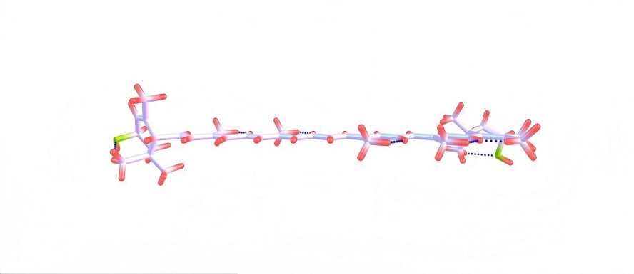
Lutein is a dihydroxy carotenoid with a symmetrical structure containing two violone rings. It has obvious lipophilic hydrophobicity. Three of the 40 carbon atoms in its structure are chiral, so there are eight isomers. Studies have found [4] that the degree to which lutein is released from different foods and utilized by the human body also varies. The degree of release of lutein and zeaxanthin from fruits such as citrus, kiwifruit, grapes, and sweet potatoes can reach 100%, while the degree of release of lutein from green leafy vegetables such as spinach and lettuce is about 38%, and the release is even better after cooking.

Because lutein contains many unsaturated structures, it has very good antioxidant properties. It can quench active oxygen free radicals, fight lipid peroxidation-induced cell degeneration, maintain the fluidity of cell membranes, and protect the body from cell damage caused by oxidative stress. It is known that the antioxidant biological activity of lutein in the diet can be 5 times higher than that of β-carotene [5]. Its antioxidant effect is achieved by the conjugated polyene chain losing one electron to form a cationic radical, which reduces oxygen free radicals, thereby inhibiting their activity and preventing damage to normal cells. In vitro studies have confirmed [5] that lutein has strong free radical scavenging capacity and antioxidant activity, The median effective concentration (EC50) for scavenging oxygen free radicals is 1.07–1.36 mg/100 ml. In a mouse model of D-galactose-induced oxidative damage, lutein at low, medium and high doses can significantly reduce the malondialdehyde level in the serum of model mice and increase superoxide dismutase activity in a dose-dependent manner [2]. In a rat model of isoproterenol-induced heart failure, lutein was given at a dose of 40 mg per day for 28 consecutive days. It was found that lutein can improve the antioxidant status of the heart by upregulating the Nrf2/HO-1 signaling pathway [2]. In addition, lutein can improve the overoxidation stress response and inflammatory response in human retinal pigment epithelial cells through the SIRT1/NLRP3 signaling pathway, thereby increasing cell survival [5].
2 Lutein in vivo process
2.1 Lutein absorption
Lutein absorption is highly dependent on dietary habits. People who eat a diet low in vegetables may have a lutein intake of less than 2 mg [6]. People who eat a diet high in vegetables may have an average lutein intake of more than 3 mg per day [7]. Due to the lipophilic nature of carotenoids, they can pass through biological membranes on their own and are more easily absorbed in a fat-soluble environment. Therefore, taking them with fatty foods can promote lutein absorption. Since egg yolk itself is rich in phospholipids, lutein in eggs is more easily absorbed by the body than lutein from other plant sources. Lutein has a high affinity for high-density lipoprotein, and 30% to 50% can be transported by high-density lipoprotein [8]. Dietary fibre, on the other hand, is not conducive to lutein absorption because it can promote the excretion of lutein in the intestine. Factors other than food can also affect lutein absorption, such as smoking, drinking, and taking medication. For example, the weight loss drug orlistat can affect fat absorption and thus inhibit lutein absorption [6].
2.2 Distribution of lutein
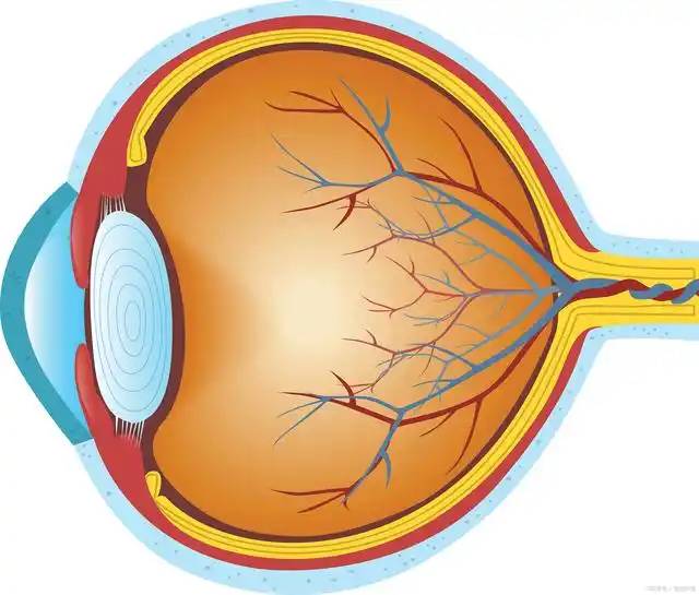
Lutein is mainly distributed in the retina, accounting for 50% to 80% of all carotenoids in the body. The concentration can reach 1 mmol/L, which is more than 1,000 times the concentration in the blood (0.1 to 1.23 μmol/L) [7]. Lutein is distributed around the fovea of the macula, mainly in the inner plexiform layer, and is also found in retinal glial cells, i.e. Muller cells. Studies have shown [7] that lutein needs to be actively transported by B1-type scavenger transporters to enter the retina, which is the physiological basis for lutein enrichment in the retina. Retinal lutein is the most important component of macular pigment, and its concentration is reflected in the macular pigment density. After three months of continuous dietary lutein intake, an increase in macular pigment density can be observed, and even after the lutein blood concentration returns to the baseline level before the three-month period, the macular pigment density can still be maintained at a relatively high level [9].
There are two theories to explain this phenomenon: one is that humans have low lutein-splitting enzyme activity, which leads to lutein accumulation; the other is that fat acts as a lutein reservoir, and lutein in fat is continuously released to maintain macular pigment density. Because in the absence of lutein supplementation, the decrease in lutein in fat cells is negatively correlated with changes in macular pigment density [9]. In addition, lutein is also distributed in the lens, and is an important substance that protects the lens from cataract damage. Lutein is most abundant in the eye during infancy, and its content decreases significantly with age [10].
3 Lutein dosage safety
According to the classification of the US Food and Drug Administration (FDA), lutein is generally safe for consumption [11]. Long-term supplementation of lutein in healthy people can help promote visual function. 115 healthy volunteers were divided into an intervention group of 57 and a control group of 58. The intervention group was given 10 mg of lutein daily for 1 year. The macular pigment density of the retina was significantly higher than that of the control group, and the color difference and light stress recovery time improved [12]; a randomized, double-blind, placebo-controlled intervention study of 120 healthy drivers for a period of 1 year found that daily supplementation with 20 mg of lutein can increase the macular pigment density of the retina in normal people and is beneficial for night driving [12].
Although most studies have found a clear dose-response relationship for lutein use, there is no statistical difference between 10 mg and 20 mg per day in terms of improving visual function if used for a long period of time. Moreover, the minimum daily dose should be 6 mg, and if it is lower than 6 mg, the protective effect on visual function will be greatly reduced [13]. Considering the different dietary habits in different regions, 10-12 mg per day should be a more appropriate dose. The recommended dose in nutrition-related studies in the United States is 20 mg per day, and the recommended dose in the Chinese Nutrition Society's “Dietary Reference Intakes for Chinese Residents” is 10 mg per day, with a tolerable upper intake level of 40 mg per day. There have also been reports of extremely rare cases of adverse reactions [14], including one elderly woman who took a lutein supplement of 20 mg per day for 8 years. Her lutein blood concentration increased to 2.9 times the normal value. The adverse reaction was mainly foveal scintillation. Seven months after discontinuing the supplement, the lens of the right eye dissolved and disappeared, and it was believed that this was related to lutein overdose.
In safety evaluation tests, extremely high doses are usually used to induce the toxicity of lutein and obtain safety data. Kruger et al. [15] gave rats 639 mg/ (kg·d) lutein orally and found no adverse reactions. Considering the differences in toxicant tolerance between different species, and using a conversion factor of 6.2 between human and rat tolerances, i.e. 40mg/(kg ·d) lutein for a 60kg person is equivalent to 4.1mg/(kg·d) for a rat, which is negligible compared to 639mg/(kg·d)[16].
4 Lutein's eye health and adjuvant therapeutic effects
4.1 Prevention and adjuvant treatment of age-related macular degeneration
Age-related macular degeneration is the leading cause of blindness from visual impairment in some countries or regions. A large number of studies have shown [13] that lutein can increase macular pigment density, visual acuity and contrast sensitivity. Research on lutein and age-related macular degeneration began in the 1990s. An additional daily lutein supplement of 10 mg for one year can effectively inhibit age-related macular degeneration and increase visual acuity and contrast sensitivity [17]. After supplementing with 20 mg of lutein per day for 3 months, the lutein dose was reduced to 10 mg per day for another 3 months, which significantly increased the macular pigment density in patients with macular degeneration [18]. For people with early macular degeneration, relevant studies have shown [15, 18] that in people aged 45 to 71, supplementing with 12 mg of lutein per day for 2 years can significantly improve visual sensitivity and reduce the risk of developing cataracts by 22% compared to the general population.
Oral vitamins and trace elements have long been used to prevent and treat age-related macular degeneration, including vitamin C, vitamin E, beta-carotene, zinc and copper supplements. Lutein, with its antioxidant properties and structural similarity to beta-carotene, is also important in preventing and treating age-related macular degeneration. In addition, macular degeneration is also a genetically related disease. The complement factor H (CFH) gene polymorphism increases the susceptibility to macular degeneration [19]. Moreover, lutein supplementation can significantly reduce the prevalence of macular degeneration in individuals carrying the CFH mutant gene, suggesting that lutein counteracts the adverse factors that lead to macular degeneration [19].
4.2 Avoid photochemical damage
With the development of the times and the needs of work, more and more people are exposed to blue light for long periods of time (such as using computers or mobile phones for several hours a day for months or years), and the number of people suffering from visual function damage as a result is increasing. This type of photochemical damage is the most common type of retinal photodamage, far more common than thermal damage or mechanical damage. Photochemical damage refers to a series of harmful chemical stimuli caused by exposure to near ultraviolet light and shorter wavelengths of visible light (400–550 nm), which can lead to cell apoptosis and retinal damage. The wavelength range of photo-chemical damage overlaps with the blue wavelengths of light sources such as neon lights, mobile phone screens, computer monitors, arc welding lights, and highland sunlight. The damage mechanism involves oxidative stress-mediated damage, damage mediated by the absorption of photons by rhodopsin, damage caused by lipofuscin-enhanced lipid peroxidation, and apoptosis. Long-term continuous blue-light exposure induces photochemical damage typical of age-related macular degeneration, accompanied by severe visual function loss.
The mechanism of lutein's protective effect against retinal photodamage mainly involves the following three items [20]. (1) Blue-light filtering: The wavelength of blue light (435–480 nm) is in the high-frequency band of visible light with high energy and great harm, and can cause damage to the retinal pigment epithelium and photoreceptor cells. At the same time, under the action of blue light, the metabolite lipofuscin in the pigment epithelium can induce the production of reactive oxygen species, leading to lipid peroxidation and apoptosis of retinal epithelial cells. The maximum absorption wavelength of lutein is 460 nm, which is within the wavelength range of blue light. It can reduce the transmission of blue light by 40% to 90%, helping to protect the retina from photochemical damage. In the outer plexiform layer of the fovea, there are rod cells and cone cells with light-sensitive receptors distributed, which is also an area rich in lutein, which can avoid blue light exposure of the light-sensitive receptors. ②Antioxidant effect: The lutein molecule is a lipid containing a continuous conjugated double bond. Its structure is similar to that of long-chain unsaturated fatty acids such as docosahexaenoic acid (DHA).
It can provide a proton to eliminate active oxygen free radicals, and after exerting its antioxidant function, it inactivates itself and is metabolized. Especially in tissues with high oxygen consumption, such as the brain and retina, which produce large amounts of aerobic metabolites during continuous work, lutein can consume these metabolites and inhibit damage to neurons and the retina. People who are exposed to continuous blue light (such as playing electronic products for a long time or programming) are often in a state of stress with continuously increasing adrenaline levels (such as staying up late). The resulting increase in oxygen consumption and elevated lipid peroxidation levels can promote the oxidation and inactivation of lutein. For people with hyperlipidemia, cholesterol and triglycerides consume lutein for esterification, resulting in a decrease in macular pigment density. ③Indirectly improves the efficiency of visual signal transmission: Some studies have found [21] that lutein can improve the survival rate of retinal cells and thus indirectly improve visual function. However, there is little relevant evidence, and further research is needed.
A study of the eye health of 2,222 undergraduate and graduate students from four universities in Beijing after long-term blue light exposure [21] analyzed the factors associated with eye symptoms and compared the relationship between blue light exposure time and eye symptoms in students from different schools and grades. The results showed that the detection rate of eye-related symptoms among college students was 66.0%, and daily blue light exposure time, mental state, and myopia were related factors affecting eye symptoms. As these students grew older, the longer their blue light exposure time, the more severe their eye-related symptoms. The highest detection rates were, in order, decreased vision, blurred vision, and dry eyes.
Volunteers were selected and given 6 mg or 12 mg of lutein daily for 12 weeks. Venous blood (5 ml) was collected in the morning on an empty stomach during the intervention period and 6 weeks after the intervention to determine serum lutein concentrations. Visual function indicators including visual acuity, contrast sensitivity, glare sensitivity, critical flicker fusion frequency, tear film break-up time, photopic persistence, and visual reaction time were also tested. The results showed that the unaided visual acuity, corrected visual acuity, and glare sensitivity of volunteers who received oral lutein 12 mg per day tended to improve. The tear film break-up time in the left eye was significantly longer than before the intervention, and the duration of bright vision was significantly improved, but there was no significant change in critical flicker fusion frequency, discrimination reaction, or choice reaction. Moreover, the serum lutein level increased rapidly after lutein intervention, suggesting that lutein has a protective effect on visual function exposed to blue light [22].
4.3 Improving myopia
Long-term reduction in macular pigment density is not only the primary cause of age-related macular degeneration, but also related to the progression of myopia, especially high myopia [23-24]. Myopia is the most common visual dysfunction in the world. Although myopia is a non-fatal disease and can be corrected with glasses and surgery, myopia itself increases the risk of other serious eye diseases, such as glaucoma, lacquer-like cracks and retinal detachment, and still needs attention.
Myopia is associated with an elongation of the eye axis. In mild myopia, the eye axis is 24 mm, and in severe myopia, it can be as long as 30 mm. Studies have shown [24] that macular pigment density is closely related to the degree of myopia. As the degree of myopia increases, the eye axis gradually becomes longer, and the macular pigment density gradually decreases. Compared with people with normal vision, myopic people with reduced macular pigment density are more likely to develop lacquer-like cracks. To ensure that images fall correctly on the retina, it is common practice to wear myopia glasses. In addition, excimer laser in situ keratomileusis or the use of orthokeratology lenses are also methods of correcting myopia. Supplementation with lutein can improve the changes in macular pigment density caused by the elongation of the myopic eye axis. A cross-sectional survey involving 4,166 subjects found [25] that the myopia incidence in people with higher blood lutein levels was lower, and in the top 20% of lutein blood concentrations, the myopia incidence was 40% lower than normal (odds ratio (OR) = 0.57, OR = 0.57, P<0.001] and the effect of lutein in improving the incidence of myopia is more significant than the possibility of ultraviolet rays causing myopia, suggesting that a higher plasma lutein level is a protective factor against myopia.
A prospective study of Japanese myopic volunteers over a period of 3 months [26] showed that after 2–3 months of oral lutein intake, the macular pigment density level in the subjects' fovea increased by 20% compared to the baseline (OR=0.725, P=0.0004), while oral zeaxanthin intake did not increase the macular pigment density in these myopic people.
In addition, another study [27] showed that lutein powder can promote the synthesis of hyaluronic acid in the eye. Hyaluronic acid, also known as hyaluronan, is a space-filling and water-retaining substance in the vitreous body of the eye. It plays an important role in light refraction. Injection of biomimetic hyaluronic acid hydrogel can control the progression of myopia in guinea pigs, and is commonly used to prevent and treat visual fatigue in people with myopia. Lutein, under the action of hyaluronic acid synthase, can activate the retinoic acid receptor and induce the synthesis of hyaluronic acid by keratinocytes, thereby improving myopia [27].
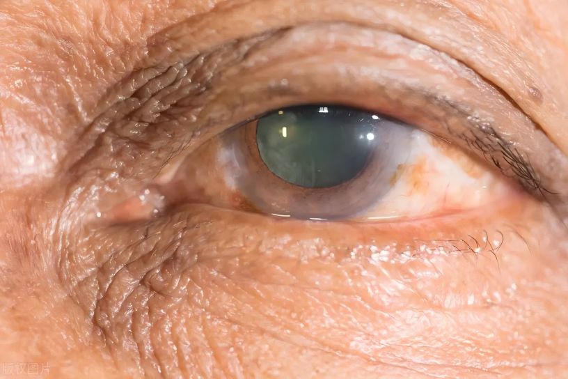
4.4 Delaying cataracts
Oxidative stress can cause damage to the lens and promote the formation of cataracts. Therefore, antioxidant therapy is also very important for preventing and treating cataracts. Lutein reduces the development of cataracts by protecting the eyes from photo-oxidative stress. Sodium selenite was used to induce cataracts in Wistar rats [28], and it was found that lutein can reduce the ratio of water-insoluble to water-soluble proteins in cataractous lenses, and relieve cataracts by regulating oxidative stress and inflammation in cataractous lenses. A study of 3,000 people with visual impairment showed [29] that the higher the blood lutein level, the lower the risk of cataracts; the more lutein consumed, the lower the risk of nuclear cataracts. High doses of lutein can reduce the risk of cataracts by 19%. For people who already have age-related cataracts, supplementing with 15 mg of lutein daily for 2 years can significantly improve the visual function of nuclear cataracts [29].
4.5 Relieving eye strain
Eye strain usually has no organic lesions and is related to genetic factors, eye habits, social environment, etc. The main symptoms are eye pain, eye swelling, dry eyes, blurred vision, etc. Affected by the pace of life and work pressure, most people, especially young students, generally spend more time looking at things and with greater intensity. Eye strain has become a common sub-health condition. A study of the relationship between lutein intake levels and symptoms of visual fatigue in college students [30] showed that 295 of the 386 college students (76%) had symptoms of visual fatigue and related eye problems, with sore eyes and vision loss being the most common. The occurrence of visual fatigue is related to computer screen light exposure, poor eye habits, and myopia. Lutein intake and outdoor activities are protective factors for the occurrence of visual fatigue, and the occurrence of visual fatigue is negatively correlated with lutein intake levels.
4.6 Reducing diabetic retinopathy
Diabetic retinopathy is the most common microvascular disease in diabetic patients, and stages I to VI are a slow and gradual process. In addition to blood glucose control, laser therapy and anti-vascular endothelial growth factor injections are the main methods for preventing blindness caused by diabetic retinopathy. Retrospective studies of optical coherence tomography and multifocal electroretinography have shown that long-term lutein or zeaxanthin supplementation can improve retinal thickness and function in diabetic patients [31-32]. Lutein and zeaxanthin levels in the plasma of diabetic patients are negatively correlated with the severity of diabetes and retinopathy. Lutein supplementation inhibits the oxidative stress, increases glutathione levels, increases the activity of glutathione peroxidase and brain-derived neurotrophic factor, and reduces the expression of various inflammatory factors. The effects of lutein supplementation include the suppression of oxidative stress, increased glutathione levels, increased activity of glutathione peroxidase and brain-derived neurotrophic factor, and reduced expression of various inflammatory factors. It also improves the survival rate of human retinal pigment epithelial cells [5] and promotes the improvement of various histological changes in the diabetic retina, such as the thickness of the inner nuclear layer, the thickness of the inner plexiform layer, and the thickness of the ganglion cell layer.
4.7 Auxiliary treatment for retinopathy of prematurity
Retinopathy of prematurity is caused by the underdeveloped retina of premature infants, which is prone to oxidative stress due to ischemia, and in turn leads to retinopathy. Retinopathy of prematurity has become the leading cause of neonatal blindness, with about 180,000 new cases of retinopathy of prematurity each year worldwide. These patients are susceptible to myopia or macular scars in adulthood. In China, the most important factor affecting retinopathy of prematurity is a gestational age of <28.6 weeks. In addition, long-term oxygen inhalation after birth, low insulin-like growth factor, cerebral ventricle hemorrhage and bloodstream infection are also factors that lead to retinopathy of prematurity [33]. Early treatment of retinopathy of prematurity can reduce visual function damage in adulthood in these people. In addition to surgery, the use of anti-vascular endothelial growth factor drugs such as bevacizumab and ranibizumab is currently the main treatment for retinopathy of prematurity. Since lutein is enriched in the retina and has the characteristic of resisting oxidative stress, timely lutein supplementation after birth has a positive effect on preventing retinopathy of prematurity.
Studies have shown [34] that lutein supplementation within 48 hours of birth can significantly reduce the amount of hydrogen peroxide in the body of the newborn and increase the body's antioxidant capacity. The incidence of retinopathy of prematurity in lutein-treated premature infants was significantly lower than that in untreated premature infants. Lutein also inhibited the progression of retinopathy in premature infants with retinopathy [35-36]. However, research on lutein as an adjuvant treatment for retinopathy of prematurity is still insufficient. The main problem is that retinopathy of prematurity is a multifactorial disease, and there is still insufficient clinical evidence to determine whether lutein deficiency can lead to retinopathy of prematurity. In recent years, some studies have reviewed the effects of lutein supplementation during pregnancy or breastfeeding on infants. It is believed that lutein, as an important substance for promoting nervous system development, suggests that adequate lutein supplementation during pregnancy is also beneficial for the prevention of retinopathy of prematurity [37].
5 Current status of lutein application fields
5.1 Lutein application in food and health food fields
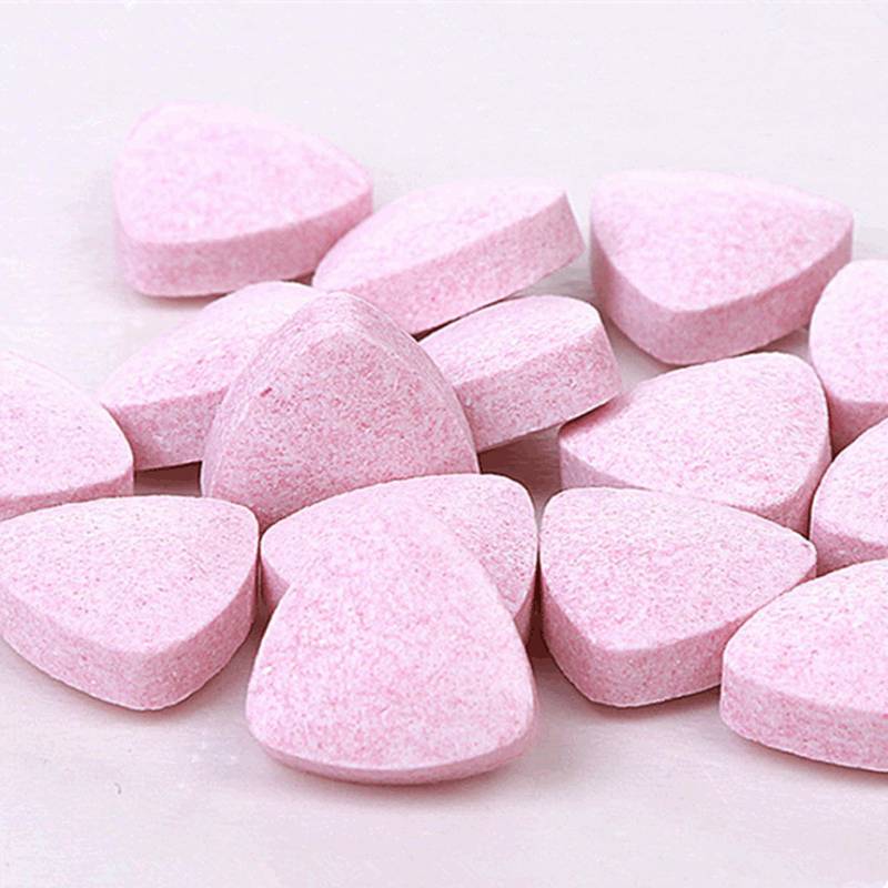
It was first noticed that natural lutein present in plants gives them a bright color and strong dyeing power. It was found that it can be used in food processing to improve the appearance and quality of food; as a feed additive, it is added to poultry feed [38-39]; and it is used to color aquatic products [40] and poultry products to improve their appearance and quality. With the deepening of research, researchers have found that lutein plays a role in eye protection. The State Food and Drug Administration has approved the marketing of lutein as one of the main active ingredients in health products that relieve eye fatigue. More and more manufacturers are producing lutein-related products, such as lutein ester tablets, blueberry lutein ester chewable tablets, Yelan Mingmu capsules, blueberry-flavored lutein ester drinks, etc., all of which contain lutein [41-44]. It can be seen that lutein is extending from food additives to the health care field and has been widely used, especially in the direction of eye health care. However, people must pay attention to the lutein content in the product during use to avoid adverse reactions caused by excessive consumption.

5.2 Application of lutein in the medical field
Currently, lutein is still under research, and there have been no reports of lutein being included in prescription drugs at home or abroad. Relevant studies have shown that lutein has a certain adjuvant therapeutic effect on ophthalmic diseases. For example, Hu Bojie et al. [43] studied the clinical application of lutein and zeaxanthin in diabetic retinopathy. For patients with simple diabetic retinopathy, there is a positive correlation between visual acuity and the serum lutein and zeaxanthin content within a certain range. Xia Liying et al. [44] conducted a clinical study on lutein treatment of age-related macular degeneration, and the results showed that the serum concentrations of lutein and zeaxanthin were positively correlated with the macular pigment concentrations in the retina.
Lutein has a certain effect on postmenopausal osteoporosis. For example, Su Yeping et al. [45] studied the effect of the combined application of lycopene, lutein and phytosterols on the intestinal flora of mice with postmenopausal osteoporosis. Lutein also plays a role in liver cancer. For example, Wang Ruozhong et al. [46] studied the inhibitory effect and mechanism of lutein on human liver cancer cells HepG2 and found that lutein reduces the level of reactive oxygen species in liver cancer cells. At present, lutein is receiving increasing attention in the medical field, and its important role is constantly being discovered, and other practical therapeutic effects are being continuously explored.
6 Summary
Lutein is a natural compound with antioxidant activity. It can accumulate in the retina and has the effect of preventing age-related macular degeneration, avoiding photochemical damage, improving myopia, delaying cataracts, relieving visual fatigue, reducing diabetic retinopathy and assisting in the treatment of retinopathy of prematurity. It is widely found in plants in nature and is already accepted as an eye health product such as blueberry lutein ester tablets and lutein eye drops. Due to its antioxidant properties and stability, researchers are exploring its potential for the prevention of cardiovascular disease, anti-osteoporosis and anti-cancer effects. It is believed that lutein will have a wider range of applications in the medical field in the near future, and it will also have good application prospects in other types of food and health food.

References
[1] Sun Wei, Yan Xiao, Li Huihua. Research progress on the relationship between phytochemicals and cardiovascular disease [J]. Chinese Food and Nutrition, 2019, 25(2): 64-67.
[2] Wang Min, Wang Xiaoli, Shen Hui. Research progress on the prevention of cardiovascular disease by lutein [J]. Occupational Health, 2020, 36(3): 424-427.
[3] Tian Li, Feng Guodong, Liu Ying, et al. Comparison of agronomic traits and lutein content of different cultivars of pigmented marigolds [J]. Journal of Hefei University of Technology (Natural Science Edition), 2018, 41(5): 703-707.
[4] GOMBAČ Z,ČRNIVEC IGO,SKRT M,et al. Stabilisation of lutein and lutein esters with polyoxyethylene sorbitan mono ole ate ,medium -chain triglyceride oil and lecithin[J] . Foods,2021,10(3):500.
[5] Wan Fang, Ma Lifang. Research on the mechanism of lutein improving the survival rate of human retinal pigment epithelial cells through the SIRT1/NLRP3 signaling pathway [J]. Tianjin Medicine, 2021, 49(2): 131-135.
[6] OLMEDILLA-ALONSO B,RODRÍGUEZ-RODRÍGUEZE,BELTRÁN-DE-MIGUEL B,et al. Changes in lutein status markers(serum and faecal concentrations,macular pigment) in response to a lutein-rich fruit or vegetable (three pieces/day)dietary intervention in normolipemic subjects[J]. Nutrients,2021,13(10):3614.
[7] LEE HS,CHO YH,PARK J, et al. Dietary intake of phytonutrients in relation to fruit and vegetable consumption in Korea[J]. J AcadNutr Diet,2013,113 (9):1194-1199.
[8] BÖHM V,LIETZ G,OLMEDILLA-ALONSO B,et al.From carotenoid intake to carotenoid blood and tissue concentrations-implications for dietary intake recommendations[J] . Nutr Rev,2021,79(5):544- 573.
[9] GIORDANO E,QUADROL. Lutein,zeaxanthin and mammalian development:metabolism,functions and implications for health[J] . Arch Biochem Biophys, 2018,647:33-40.
[10] GAZZOLO D,PIC ONE S,GAIERO A,et al. Early pediatric benefit of lutein for maturing eyes and brain-an overview[J] . Nutrients,2021,13(9):3239.
[11] RANARD KM,JEON S,MOHNES, et al. Dietary guidance f or l utein:con s ide rati on f or in take recommendations is scientifically supported[J] . Eur J Nutr,2017,56(Suppl 3):37-42
[12] Zheng Ying, Chen Xinbin, Ma Yan, et al. Research progress on the biological function of lutein and related chronic diseases [J]. Chinese Food and Nutrition, 2023, 29 (3): 51-55, 10.
[13] WILSON LM,THARMARAJAH S,JIA YX,et al. The effect of lutein/zeaxanthin intake on human macular pigment optical density:a systematic review and meta analysis[J] . Adv Nutr,2021,12(6):2244-2254.
[14] CHOI RY,CHORTKOFF SC,GORUSUPUDIA,et al.Crystalline maculopathy associated with high-dose lutein supplementation[J] . JAMA Ophthalmol,2016,134 (12):1445-1448.
[15] KRUGER CL,MURPHY M ,DEFREITAS Z,et al. Aninnovative approach to the determination of safety for a dietary ingredient derived from a new source:case study using a crystalline lutein product[J]. Food Chem Toxicol,2002,40(11):1535-1549.
[16] NAIR AB,JACOB S. A simple practice guide for dose conversion between animals and human[J]. J Basic Clin Pharm,2016,7(2):27-31
[17] MROWICKA M,MROWICKI J,KUCHARSKA E,et al. Lutein and zeaxanthin and their roles in age-related macular degeneration-neurodegenerative disease[J]. Nutrients,2022,14(4):827.
[18] FENG LW,NIE KL,JIANG H,et al. Effects of lutein supplementation in age-related macular degeneration [J] . PLoS One,2019,14(12):e0227048.
[19] TOOMEY CB,JOHNSON LV,BOWES RICKMAN C. Complement factor H in AMD:bridging genetic associations and pathobiology[J] . Prog Retin Eye Res, 2018,62:38-57.
[20] CHAE SY,SHIN MC,JEON S,et al. A simple route to the complexation of lutein with reduced graphene oxide nanocarriers and antioxidant protection against blue light [J] . IntJ Nanomedicine,2021,16:6843-6860.
[21] Wan Fang, Ma Lifang. Research on the mechanism of lutein improving the survival rate of human retinal pigment epithelial cells through the SIRT1/NLRP3 signaling pathway [J]. Tianjin Medicine, 2021, 49(2): 131-135.
[22] Ma L. Investigation of eye-related symptoms in college students exposed to computer screens and the effect of lutein intervention on visual function and changes in serum lutein [D]. Beijing: Peking University, 2009.
[23] Qi Y, Hao J, Zhang Y, et al. Study on macular pigment optical density in highly myopic eyes [J]. Beijing Medical Journal, 2021, 43(11): 1110-1112.
[24] Liu Jue, Zhang Jing, Xu Ying, et al. The relationship between macular pigment density and thickness and myopia in adults [J]. Chinese Journal of Optometry and Vision Science, 2019, 21(12): 924-929.
[25] WILLIAMS KM,BENTHAM GC,YOUNG IS,et al. Association between myopia,ultraviolet B radiation exposure,serum vitamin D concentrations,and genetic polymorphisms in vitamin D metabolic pathways in a multicountry European study[J] . JAMA Ophthalmol, 2017,135(1):47-53.
[26] TANITO M,OBANA A,GOHTO Y, et al. Macular pigment density changes in Japanese individuals supplemented with lutein or zeaxanthin:quantificationvia resonance Raman spectrophotometry and autofluorescence imaging[J]. Jpn J Ophthalmol,2012,56(5):488-496.
[27] Yin Chunjie, Gan Qian, Zhang Qian. Nutrients and myopia [J]. Health Research, 2022, 51(5): 720-724.
[28] Wei Yiyang, Ma Bowei, Li Hailong, et al. Research progress in the treatment of cataracts with traditional Chinese medicine monomers [J]. Chinese Journal of Gerontology, 2022, 42(17): 4387-4391.
[29] LIU XH,YU RB,LIU R,et al. Association between lutein and zeaxanthin status and the risk of cataract:a meta-analysis[J] . Nutrients,2014,6(1):452-465.
[30] Yang Linjuan, Zhang Xiuli, Liang Bin. A study on the correlation between lutein intake levels and symptoms of visual fatigue in college students [J]. China Food and Nutrition, 2019, 25(6): 87-89.
[31] MOSCHOS MM,DETTORAKI M,TSATSOS M,et al. Effect of carotenoids dietary supplementation on macular function in diabetic patients[J]. Eye Vis,2017,4:23.
[32] PAN FH,CUI WX,GAO L,et al. Serum lutein is a promising biomarker for type 2 diabetes mellitus and diabetic kidney disease in the elderly[J] . J Clin Lab Anal,2022,36(4):e24350.
[33] Wang Y, Wu S, Xiao Y. New progress in the study of risk factors for retinopathy of prematurity [J]. Chinese Journal of Strabismus and Pediatric Ophthalmology, 2022, 30(3): 47-48.
[34] GRAZIOSI A,PERROTTA M,RUSSO D,et al. Oxidative stress markers and the retinopathy of prematurity[J] . J Clin Med,2020,9(9):2711.
[35] RUBIN LP,CHAN GM,BARRETT-REIS BM,et al. Effect of carotenoid supplementation on plasma carotenoids, inflammation and visual development in preterm infants [J] . J Perinatol,2012,32(6):418-424.
[36] MANZONI P,GUARDIONE R,BONETTI P,et al. Lutein and zeaxanthin supplementation in preterm very low- birth-weight neonates in neonatal intensive care units: a multicenter randomized controlled trial[J] . Am J Perinatol,2013,30(1):25-32.
[37] CHRISTIFANO DN,CHOLLET-HINTON L,HOYER D, et al. Intake of eggs,choline,lutein,zeaxanthin,and DHA during pregnancy and their relationship to fetal neurodevelopment[J] . Nutr Neurosci,2023,26(8): 749-755.
[38] Xu Lancheng, Yang Shengkun, Wang Chenqin, et al. Research on the biological activity of marigold and its application in animal production [J]. Feed Research, 2022, 45(2): 141-144.
[39] JUAN-LUIS F,ZAIDA M,MARA C,et al. Outdoor large-scale cultivation of the acidophilic microalga Coccomyxa onubensis in a vertical close photobioreactor for lutein production[J]. Processes,2020,8(3):324.
[40] Lv Yong, Ye Jiaping, Peng Fuyou, et al. Effect of natural lutein and compound xanthophyll on the coloring effect of yellow-breasted broilers [J]. Guangdong Feed, 2020, 29(10): 31-34.
[41] Li Xiang, Sun Siqi, Liu Xiaoyun, et al. Research on the preparation process and determination of the content of effective ingredients in the health food Ye Lan Mingmu capsule [J]. Food and Drugs, 2022, 24(4): 314-318.
[42] Sun Zhangqing, Jiang Jingjing, Zhou Li, et al. Development of a blueberry-flavored lutein ester drink [J]. Food Safety Herald, 2023(18): 108-110.
[43] Hu Bojie, Hu Yanan, Lin Song, et al. Clinical application of lutein and zeaxanthin in diabetic retinopathy [J]. New Advances in Ophthalmology, 2010, 30(9): 866-868.
[44] Xia Liying, Liu Weijia, Kou Qiuai, et al. Clinical study of lutein in the treatment of age-related macular degeneration [J]. World Traditional Chinese Medicine, 2013, 8(5): 517-519.
[45] Su Yeping, Xie Peiya, Wei Ruifei, et al. The effect of the combined application of lycopene, lutein and phytosterols on the intestinal flora of postmenopausal osteoporosis mice [J]. Journal of Guangxi Medical University, 2024, 41(3): 388-397.
[46] Wang RZ, Shen XN, Shi DY, et al. Lutein inhibits human hepatoma HepG2 cells and its mechanism. Journal of Nutrition, 2012, 34(4): 332-335.


