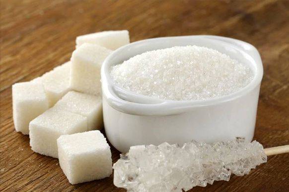
What Is Sweetener D Tagatose?
With the increasing demand for a better quality of life, the types of food and beverage sweeteners are constantly increasing. In addition to nutritive sweeteners such as sucrose, non-nutritive sweeteners are becoming the mainstream of the sweetener market because they rarely or do not produce calories, and can reduce the risk of diabetes, obesity, cardiovascular disease and cancer. Currently commonly used non-nutritive sweeteners include fructooligosaccharides, erythritol, xylitol, etc. D-tagatose, discovered in recent years, is a sweetener with special health benefits and has great market potential.
1 Introduction to D-tagatose
D-tagatose (CAS 87-81-0) is a rare natural hexose that exists in nature. It is a diastereoisomer of fructose with a relative molecular mass of 180.16. It has a sweetness similar to sucrose, with a sweetness level of 92% of sucrose, and has virtually no unpleasant aftertaste or off-flavors. It produces only 1/3 of the calories of sucrose, making it a low-calorie sweetener. It is estimated to have a glycemic index of 8.
Tagatose can be metabolized via the tagatose-6-phosphate pathway, which is present in some microorganisms but not in higher animals. Only about 20% of the tagatose consumed by humans is absorbed in the small intestine, and the rest is selectively broken down and utilized by intestinal microorganisms [1]. Therefore, tagatose is low in calories and has the effect of controlling weight and preventing diabetes [2]. Tagatose produces low levels of acid in the mouth and does not lower the pH of dental plaque, effectively preventing enamel erosion and the occurrence of dental caries [3]. Tagatose is also a good prebiotic. Toxicological tests have shown that tagatose is safe and non-toxic [4]. The health functions and application fields of tagatose are shown in Table 1.
2 Mechanism of tagatose against hyperglycemia
The mechanism of tagatose against hyperglycemia has not yet been fully explained. Based on experimental studies, a possible mechanism for the control of blood glucose concentration by tagatose has been proposed. After absorption, tagatose is mainly metabolized in the liver, and the metabolic pathway is the same as that of fructose, that is, fructokinase is first phosphorylated to tagatose-1-phosphate, and then aldolase is broken down into glyceraldehyde and dihydroxyacetone phosphate, and the decomposition rate is about half that of fructose-1-phosphate. Similar to fructose-1-phosphate, elevated concentrations of tagatose-1-phosphate stimulate the activity of glucose kinase, resulting in an increased level of glucose phosphorylation to gluconate-6-phosphate and further activation of glycogen synthase. At the same time, tagatose-1-phosphate and fructose-1-phosphate inhibit glycogen phosphorylase [5]. The overall effect of these enzymes on glycogen synthesis and glycogen breakdown is a decrease in blood glucose (Figure 1). As can be seen in Figure 1, fructose has a similar mechanism to tagatose for lowering blood glucose, but tagatose is more effective [6] and has fewer side effects [7].
3 Application of tagatose
In 2001, the US FDA determined that tagatose was Generally Recognized as Safe (GRAS) [8]. In July of the same year, the Joint Expert Committee on Food Additives (JECFA) recommended tagatose as a new low-calorie sweetener that could be used as a food additive with an ADI (acceptable daily intake) of 0–80 mg/(kg•d). Subsequently, tagatose began to be widely used in healthy drinks, yogurt, fruit juice, foods for diabetics, diet foods, chewing gum, cereal foods, meat products, candy, etc., and in medicine for cough syrup, powders, effervescent agents, adhesives for fixing dentures, and oral disinfectants.
In 2003, PepsiCo began using tagatose in Sprite drinks, marking the first time tagatose had been used in a commercial product. Subsequently, New Zealand's Miada Sports Nutrition Foods Company used tagatose in chocolate products, which were launched in the Australian and New Zealand markets in May 2003.

Research into tagatose as a treatment for diabetes has already begun. On November 16, 2009, Spherix reported that the Phase III clinical trial had achieved the expected results. The company will submit a new drug application in 2010 to use tagatose to treat type II diabetes [9].
4 Tagatose production
There are two methods for producing tagatose: chemical synthesis and biotransformation. Chemical synthesis uses a soluble alkali metal salt or alkaline earth metal salt as a catalyst to promote the formation of tagatose from D-galactose under alkaline conditions, and the formation of a metal hydroxide-tagatose complex, and then neutralization with acid to obtain D-tagatose [10]. Because chemical methods are energy intensive, the products are complex, purification is difficult, there are many side reactions, and chemical pollution is produced, the bioconversion method has better application prospects.
At present, the method of biotransformation for the production of tagatose that has been studied more is the use of L-arabinose isomerase to catalyze the conversion of D-galactose to tagatose. The natural function of L-arabinose isomerase (EC 5.3.1.4, L-arabinose isomerase, L-AI) is to catalyze the production of L-ribulose from L-arabinose. In recent years, it has been discovered that this enzyme can also catalyze the conversion of D-galactose to tagatose. The L-AI coding gene is widely found in prokaryotes, including Acidothermus cellulolyticus (Acidothermus cellulolytics) [11], Alicyclobacillus acidoc aldarius, whose source AI is hereinafter referred to as AAAI) [12], Geobacillus stearothermophilus (GSAI) [13, 14], Thermoanaerobacter mathranii [1 5], Thermotoga maritima [16], Thermotoga neapolitana [17], Thermus sp. IM6501 [18], etc. It has been found that the optimum temperature range for L-AI is 20~ 80 ℃, and the optimum pH (pHopt) is 6.0~ 8.0. Metal ions such as Mn2+ or Co2+ can improve its stability. There is still much room for improvement in the industrial production of L-AI. The main research directions for the catalytic production of tagatose using L-AI are described below.
4.1 Improving the catalytic activity of the enzyme
The specific activity of L-arabinose is about 30 U/mg, while that of L-arabinose is less than 10 U/mg. Initial attempts have been made abroad to improve the substrate affinity and catalytic efficiency of L-AI through enzyme engineering. Kim et al. [19] randomly mutated and directed modified GSAI. In the first round of evolution, there were five amino acid site mutations, and the specific activity of the enzyme increased 11 times; in the second round, there were three more site mutations, and the specific activity of the enzyme increased another 5 times [20]. Among the eight mutant sites, the A408V and K475N mutations have a significant impact on the enzyme's substrate affinity and reaction rate. Recently, researchers have identified two L-AI with extremely strong substrate specificity from Bacillus licheniformis ATCC 14580 and B. subtilis str. 168, which only use L-arabinose as a substrate [21-22]. By comparing their primary sequences with other L-AI and performing advanced structural analysis, it is hoped that the rules governing the substrate specificity of L-AI can be revealed, thus providing a basis for enzyme engineering.
4.2 Reducing the optimum pH of the enzyme
The pHopt of L-AI is mostly in the alkaline range, while industrial conversion is more suitable in the acidic range because (1) tagatose is stable at pH 2–7, and high pH values increase side reactions; (2) lactose is usually used as a raw material in production to hydrolyzing lactose to produce galactose, and then isomerizing galactose to tagatose. Lactose hydrolysis is usually carried out under acidic conditions (pH 5.0~6.0), and the use of acidic L-AI can simplify the process and save costs.
The first way to obtain acidic L-AI is to screen various microorganisms, especially acidophilic microorganisms, such as Bacillus acidopullulyticus (pH opt 6.0, 65 ° C, ATCC 43030) [12]. There are also some L-AI derived from acidophilic bacteria whose pHopt is in the neutral range, but which can maintain most of their activity and maintain a certain stability under acidic conditions, such as L-AI derived from Acidithiobacillus ferrooxidans [11].
Another way to obtain acidic L-AI is enzymatic engineering. Lee et al. [12, 23] compared the amino acid sequences of AAAI (pHopt = 6), GSAI (pHopt = 7) and BHAI (from B. halodurans, pHopt = 8) and proposed that 269 lysine residues were determined to be responsible for the acid adaptation of AAAI. Subsequent site-directed mutagenesis confirmed this hypothesis: the pHopt of AAAI-K269E shifted by one pH unit to the alkaline range, while the pHopt of BHAI-E268K shifted by one unit to the acidic range. Oh et al. [24] performed site-directed random mutagenesis on Val408 and Asn475 of another GSAI (pHopt = 8.5), obtaining two mutants with a pHopt of 7.5, Q408V and R408V. Recently, Rhimi et al. [25] rationally engineered L-AI (BSAI) from B. stearothermophilus US100. One mutant, Q268K, had better acid resistance than the wild-type enzyme, which is consistent with the results of Lee et al. It can be seen that the change in the optimum pH of L-AI can be achieved by mutating one or more amino acid sites. At present, it can be determined that the amino acid sites that affect pHopt include Lys269 (AAAI, corresponding to Glu268 of BHAI and Gln268 of BSAI) and Val408 (GSAI). Double mutation of the above two sites and the identification of other amino acid sites that affect pHopt are possible paths for further acidification of L-AI in the future.
4.3 Achieving metal ion-independent thermal stability
The reaction temperature has a significant effect on the galactose isomerization reaction. As the temperature increases, the binding reaction ability of L-AI to galactose increases, the reaction rate accelerates, the equilibrium point shifts towards the product, and the conversion rate increases significantly. However, a temperature higher than 80 °C will lead to browning, so the suitable reaction temperature for industrial production is 60-70 °C. Some thermophilic or extremely thermophilic bacteria, such as T. mathranii[15], G. stearothermophilus[23], G. thermodenitrificans[26], B. stearothermophilus US100[13] and Thertmus sp.[18], have been cloned and identified as L-AI with an optimal reaction temperature and good thermal stability.
However, these L-AI depend on metal ions to maintain their thermal stability. Since the metal ions added during the reaction need to be removed from the product, this adds cost and creates pollution. Therefore, a metal ion-independent thermally stable L-AI is needed. An L-AI from B. stearothermophilus US100 is metal ion independent below 65 °C and does not require Mn2+ to maintain enzyme activity above 65 °C, although stability is affected. Only 0.2 mmol/L Co2+ and 1 mmol/L Mn2+ are required to maintain the thermal stability of the protein [13]. It is possible to obtain an L-AI that is completely independent of metal ions through further mutation and screening.
In addition, the crystal structure of L-AI derived from Escherichia coli has been determined [27], and the three-dimensional structure of L-AI from various sources can be constructed through molecular simulation, which will provide a theoretical basis for enzyme engineering research.
4.4 Production and immobilization of enzyme preparations
At present, the heterologous expression of L-AI is mostly carried out in Escherichia coli. However, since tagatose is used in food and medicine, its expression in non-food-safe microorganisms may pose safety problems. Therefore, after the engineering of the enzyme to obtain L-AI suitable for industrial production applications, the next step is to express it in food-safe microorganisms. Commonly used food-safe microorganisms include Bacillus, Corynebacterium and yeast.
After obtaining engineered bacteria with high L-AI expression, biocatalysts can be prepared using immobilized enzymes or immobilized cells. Currently, the sodium alginate-calcium chloride method is mostly used for immobilizing L-AI enzyme preparations. However, borrowing the methods of immobilized enzymes and immobilized cells that are currently successfully used in industry can further improve efficiency, increase half-life and reduce costs.
5 Outlook
Tagatose has been discovered for several decades, and foreign researchers have conducted relatively thorough research on its physical and chemical properties and physiological functions. It has been used as a food and pharmaceutical additive in the production of products in many countries, and relevant standards and regulations have been formulated. Business Consulting (BBC) predicts that the market share of tagatose will show a clear upward trend. There has been little research on tagatose in China, and only in recent years has there been some related research. Industrial production and application of tagatose has not yet been reported, and the market for tagatose consumption is still blank. Therefore, there is huge potential for domestic production and application of tagatose, waiting to be developed by domestic researchers.
Reference
[1] Normen L, Laerke H N, Jensen B B, et al. Small-bowel absorption of D-tagatose and related effects on carbohydrate digestibility: an ileostomy study[J]. Am J Clin Nutr, 2001, 73( 1): 105-110.
[2] Donner TW, Wilber J F, Ostrowski D. D-tagatose, a novel hexose: acute effects on carbohydrate tolerance in subjects with and without type 2 diabetes[J]. Diabetes Obes Metab, 1999, 1(5): 285- 291.
[3]Wong D. Sweetener determined safe in drugs, mouthwashes, and toothpastes[J]. Dent Today, 2000, 19(5): 32, 34-35.
[4] Levin G V. Use of tagatose to enhance key blood factors: US, 6015793[P]. 2000-01-18.
[5] Ercan-Fang N, Gannon M C, Rath V L, et al. Integrated effects of multiple modulators on human liver glycogen phosphorylase a[J].Am JPhysiolEndocrinol Metab, 2002, 283( 1): E29-37.
[6]Buemann B, Toubro S, Holst J J, et al. D-tagatose, a stereoisomer of D-fructose , increases blood uric acid concentration[J] . Metabolism, 2000, 49(8): 969-976.
[7]American Diabetes Association . Evidence-based nutrition principles and recommendations for the treatment and prevention of diabetes and related complications[J]. Diabetes Care, 2002, 25( 1): 202-212.
[8]Food and Drug Administration, HHS., US. Food labeling: health claims; D-tagatose and dental caries. Final rule[J]. Fed Regist, 2003, 68( 128): 39831-39833.
[9]Spherix, Incorporated. D-tagatose[OL]. [2010-03-18]. http://www. spherix.com/ biospherics _d-tagatose.html.
[10] Beadle J R, Saunders J P, Wajda Jr. T J. Process for manufacturing tagatose: US, 5078796[P]. 1992-01-07.
[11] Cheng L, Mu W, Zhang T, et al. An L-arabinose isomerase from Acidothermus cellulolytics ATCC 43068: cloning, expression, purification, and characterization[J]. Appl Microbiol Biotechnol,2009.
[12] Lee S J , Lee D W, Choe E A , et al. Characterization of a thermoacidophilic L-arabinose isomerase from Alicyclobacillus acidocaldarius: role of Lys-269 in pH optimum[J]. Appl Environ Microbiol, 2005, 71( 12): 7888-7896.
[13] Rhimi M , Bejar S. Cloning , purification and biochemical characterization of metallic-ions independent and thermoactive l-arabinose isomerase from the Bacillus stearothermophilus US100 strain[J]. Biochim Biophys Acta, 2006, 1760(2): 191-199.
[14] Jung E S, Kim H J, Oh D K. Tagatose production by immobilized recombinant Escherichia coli cells containing Geobacillus stearothermophilus L-arabinose isomerase mutant in a packed-bed bioreactor[J]. Biotechnol Prog, 2005, 21(4): 1335-1340.
[15] Jorgensen F, Hansen O C, Stougaard P. Enzymatic conversion of D-galactose to D-tagatose: heterologous expression and characterisation of a thermostable L-arabinose isomerase from Thermoanaerobacter mathranii[J]. Appl Microbiol Biotechnol, 2004, 64(6): 816-822.
[16] Lee D W, Jang H J, Choe E A, et al. Characterization of a thermostable L-arabinose (D-galactose) isomerase from the hyperthermophilic eubacterium Thermotoga maritima[J]. Appl Environ Microbiol, 2004, 70(3): 1397-1404.
[17] Kim B C, Lee Y H, Lee H S, et al. Cloning, expression and characterization of L-arabinose isomerase from Thermotoga neapolitana: bioconversion of D-galactose to D-tagatose using the enzyme[J]. FEMS MicrobiolLett, 2002, 212( 1): 121-126.
[18] Kim J W, Kim Y W, Roh H J, et al. Production of tagatose by a recombinant thermostable L-arabinose isomerase from Thermus sp. IM6501[J]. BiotechnolLett, 2003, 25( 12): 963-967.
[19] Kim P, Yoon S H, Seo M J, et al. Improvement of tagatose conversion rate by genetic evolution of thermostable galactose isomerase[J]. BiotechnolAppl Biochem, 2001, 34(Pt 2): 99-102.
[20]Oh H J, Kim H J, Oh D K. Increase in D-tagatose production rate by site-directed mutagenesis of L-arabinose isomerase from Geobacillus thermodenitrificans[J]. Biotechnol Lett, 2006, 28(3):145-149.
[21] Kim J-H , Prabhu P, Jeya M , et al. Characterization of an L-arabinose isomerase from Bacillus subtilis[J]. Appl MicrobBiot, 2009, 85(6): 1839-1847.
[22]Prabhu P, Tiwari M K, Jeya M, et al. Cloning and characterization of a novel L-arabinose isomerase from Bacillus licheniformis[J]. Appl Microbiol Biotechnol, 2008, 81(2): 283-290.
[23] Lee D W, Choe E A, Kim S B, et al. Distinct metal dependence for catalytic and structural functions in the L-arabinose isomerases from the mesophilic Bacillus halodurans and the thermophilic Geobacillus stearothermophilus[J]. Arch Biochem Biophys, 2005, 434(2): 333-343.
[24]Oh D K, Oh H J, Kim H J, et al. Modification of optimal pH in L-arabinose isomerase from Geobacillus stearothermophilus for D-galactose isomerization[J]. J Mol Catal B - Enzym, 2006, 43( 1-4): 108-112.
[25]Rhimi M, Aghajari N, Juy M, et al. Rational design of Bacillus stearothermophilus US100 L-arabinose isomerase: potential applications for D-tagatose production[J]. Biochimie, 2009, 91(5):650-653.
[26]Kim H J , Oh D K. Purification and characterization of an L-arabinose isomerase from an isolated strain of Geobacillus thermodenitrificans producing D-tagatose[J]. J Biotechnol, 2005, 120(2): 162-173.
[27]Manjasetty B A, Chance M R. Crystal structure of Escherichia coli L-arabinose isomerase (ECAI) , the putative target of biological tagatose production[J]. J Mol Biol, 2006, 360(2): 297-309.
-
Prev
How to Get Enzymatically Modified Stevia Glucosyl Stevia?
-
Next
What Are the Uses of Sweetener D Tagatose in the Food Field?


 English
 French
 Spanish
 Russian
 Korean
 Japanese


