

 English
 French
 Spanish
 Russian
 Korean
 Japanese
What Is β 1 3 Glucan?
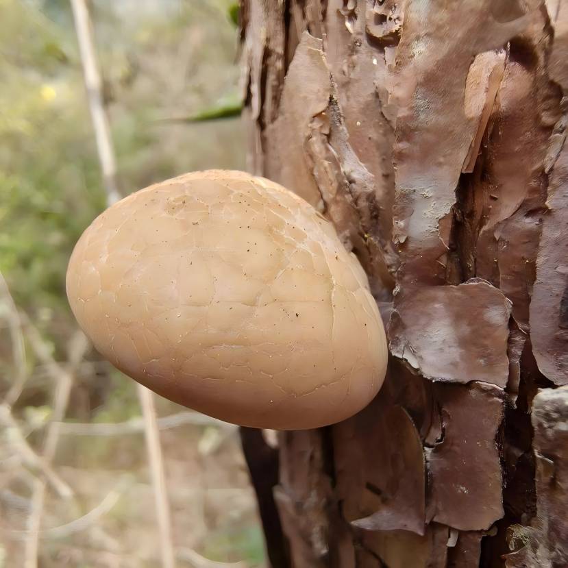
Beta-1,3-glucan is a class of high-molecular polysaccharides widely found in nature, with a main chain of glucose units linked by beta-1,3-glycosidic bonds. Some natural β-1,3-glucans also contain side chains linked by β-1,6-glycosidic bonds in varying proportions and sizes. For example, the side chains of fucoidan from brown algae contain about 30% β-1,6-linked branched structures and are therefore water-soluble [1]. Natural β-1,3-glucans are widely distributed in fungi, bacteria and plants. Common β-1,3-glucans include fucoidan, carrageenan, yeast glucan, poria cocos polysaccharide, shiitake mushroom polysaccharide, callose, etc. Due to the way in which the β-1,3-glycosidic bonds are connected and the hydrogen bond interactions between the molecules, long-chain β-1,3-glucans usually exhibit different helical tertiary structures in their natural state [2]. These special tertiary structures endow β-1,3-glucans with diverse biological functions, including regulating immunity [3], promoting the proliferation of intestinal probiotics [4], regulating blood sugar balance and lowering cholesterol [5 ]. The biological activity of β-1,3-glucan and its special tertiary structure have attracted widespread attention in the fields of food, daily chemicals, and medicine.
β-1,3-glucanase is a class of enzymes that can hydrolyze glucan linked by β-1,3-glycosidic bonds, and plays an important role in the biodegradation, reconstruction, and development and application of β-1,3-glucan. In nature, β-1,3-glucanase is widely distributed in archaea, bacteria, fungi, higher plants and animals. β-1,3-glucanase has a wide range of applications in the food and pharmaceutical industries, such as the preparation of low molecular weight β-1,3-glucan oligosaccharides, reducing the viscosity of beer fermentation broth, and inhibiting post-harvest pathogenic fungi in fruits and vegetables.

Although researchers have done a lot of research on the expression and purification, molecular structure, enzymatic properties, and catalytic mechanism of β-1,3-glucanase, the complex structure of different types of β-1,3-glucan limits its wide application. The synergistic participation of β-1,3-glucanases from different families and with different modes of action is required for the efficient degradation of complex β-1,3-glucans. Therefore, a thorough understanding of the structure, function, and catalytic mode of different types of β-1,3-glucanases is of great significance for the application and further molecular modification of β-1,3-glucanases. This paper summarizes the research progress in the structure, function and application fields of typical β-1,3 glucanases, with the aim of providing a reference for subsequent research on their catalytic mechanisms and their application in the fields of food, medicine and other fields.
1 Classification of β-1,3 glucan and their catalytic mechanisms
β-1,3 glucanases can be divided into endo-type and exo-type according to their catalytic mode. Endo-β-1,3-glucanase (EC 3.2.1.39), also known as kombu polysaccharide enzyme, is an enzyme that specifically hydrolyzes the β-1,3-glycosidic bonds in the β-1,3-glucan sugar chains. It plays a key biological role in the decomposition and reconstruction of β-1,3-glucan in nature and is family. Endo-1,3-β-glucanases hydrolyze β-1,3-glucan from the inside of the sugar chain, producing a series of oligosaccharides with different degrees of polymerization. Exo-β-1,3-glucanase (EC 3.2.1.58) hydrolyzes β-1,3-glucan substrates one after the other from the non-reducing end of the sugar chain. The hydrolyzed products are generally glucose or single oligosaccharides, and they play a supporting role in the degradation of β-1,3-glucan. Based on their evolutionary relationship, the discovered β-1,3-glucanases can be classified into 12 glycoside hydrolase (GH) families according to the CAZy database (http:// www.cazy.org/). Among them, endo-β-1,3-glucanases can be classified into 9 GH families (Figure 1): GH16, GH17, GH55, GH64, GH81, GH128, GH152, GH157 and GH158; exo-β-1,3-glucanases that have been discovered can be classified into 6 GH families: GH3, GH5, GH17 , GH55, GH128 and GH132.

β-1,3-glucanases have two hydrolysis mechanisms, namely the conservative mechanism and the inverting mechanism [6]. The catalytic process of the retained hydrolysis mechanism is divided into two steps. First, the glutamic acid residue in the active center acts as a general acid (donor of electrons) to provide a proton to the oxygen of the glycosidic bond to remove the leaving group, break the β-1,3-glycosidic bond, and form an enzyme-glycoside intermediate.
Subsequently, the glutamic acid residue in the active center acts as a broad base, assisting the water molecule in attacking the heteroatom carbon position of the enzyme-glycoside intermediate. Finally, the β-1,3-glycosidic bond of the substrate is hydrolyzed, forming the final hydrolytic product. The retention mechanism is so called because the conformation of the substrate is retained by the two inversions at the heteroatom carbon. The inverting mechanism of β-1,3-glucanase involves two conserved catalytic residues, a general acid and a general base. During the reaction, the general acid first donates a proton to the anomeric carbon of the substrate, while the general base removes a proton from a water molecule, increasing its nucleophilicity and promoting its attack on the center of the anomeric group, thereby breaking the glycosidic bond and generating the hydrolytic product (Figure 2).
2. Sources and preparation of 1,3-glucan
β-1,3-glucan is mainly derived from fungi, bacteria, plants, insects and molluscs. Table 1 summarizes the typical β-1,3-glucanases from different sources that have been reported to have application potential. The main bacteria that have been reported to produce β-1,3-glucanase are Pyrococcus furiosus [7], Paenibacillus polymyxa [8], Bacillus lehensis G1 [9], Streptomyces sp. [10], etc. Fungi and plants are also important sources of β-1,3-glucanase, including Aspergillus fumigatus [11], Trichoderma asperellum [12], Phanerochaete chrysosporium [13], barley [14], grapes [15], etc.
Natural sources of β-1,3-glucans are abundant, stable in nature, and exhibit specific catalytic activity, showing unique application potential. For example, β-1,3-glucanase produced by Paenibacillus terrae [16] can effectively inhibit the growth of plant pathogenic fungi and play an important role in plant protection. β-1,3-glucanase derived from Arca inflata [17] has β-1,3-glucanase has high activity and exhibits an immune-enhancing effect on tumor necrosis factor.
β-1,3-glucan from Trichoderma harzianum [18] is an ideal candidate enzyme for the production of β-1,3-oligosaccharides and can be used for the industrial preparation of oligosaccharides. In addition to discovering β-1,3-glucanases with excellent properties from natural sources, heterologous recombinant expression technology can also be used to discover and prepare β-1,3-glucanases, thereby expanding their sources, increasing expression levels, and expanding the scope of β-1,3-glucanase applications.
At present, the hosts for the recombinant expression of β-1,3-glucan mainly include Escherichia coli, Pichia pastoris, Bacillus subtilis, etc. The E. coli expression system is more technologically mature and relatively simple to operate than the yeast expression system, and is widely used in the discovery and preparation of novel β-1,3-glucan. For example, β-1,3-glucan from sources such as the antagonistic yeast Pichia guilliermondii [19], the macrogenome of moose rumen microorganisms [20], and Pseudomonas aeruginosa [21] are all obtained by recombinant preparation in E. coli. enzymes are all obtained by recombinant preparation of Escherichia coli. The β-1,3-glucan MoGluB derived from Magnaporthe oryzae can be efficiently expressed by the E. coli system and exhibits antifungal activity [22].
Although prokaryotic expression in E. coli has the advantages of rapid growth and low cost, some eukaryotic genes cannot be expressed effectively because the prokaryotic expression system cannot recognize eukaryotic transcription and translation elements and does not have the function of post-translational modification. Therefore, in recent years, researchers have also commonly used eukaryotic expression systems to express β-1,3-glucanase, such as Pichia pastoris and Bacillus subtilis expression systems [23]. During the inducible expression process of Pichia pastoris, the amount of protein secreted by itself is relatively small, so the expression amount of the target protein is relatively high.
However, because some β-1,3-glucan have a hydrolytic effect on the yeast cell wall, their applicability needs to be further verified experimentally. Heterologous recombinant expression technology has now become an important method for the research and preparation of β-1,3-glucan applications. Meanwhile, combining structural biology, molecular biology, directed evolution and other research methods to molecularly modify the β-1,3-glucanobtained by heterologous recombination can further improve the catalytic performance and application value of the enzyme. Feng Jianwei et al. [24] discovered a thermophilic β-1,3-glucan from compost. By site-directed mutagenesis, the 160 amino acids were changed from lysine to glutamic acid, which increased its enzyme activity by 1 7%. Muhammed et al. [25] used the modeller and I-TASSER programs to perform homology modeling of a β-1,3-glucan from the yeast Wickerhamomyces normus NCYC 434. Subsequently, the thermal stability of the model was enhanced using SPDBViewer and AUTO-MUTE. It was found that the mutant E186R had the best thermal stability, and its melting temperature was increased by 9.58 K.
3 Structure and catalytic mechanism of endo-1,3-glucan
3.1 Endo-β-1,3-glucan
Endo-β-1,3-glucan, also known as kombu polysaccharide enzyme, can specifically hydrolyze and cut β-1,3-glycosidic bonds at random from the inside of β-1,3-glucan chains, producing oligosaccharides of varying lengths. It has been reported that endo-β-1,3-glucanases are mainly distributed in the four GH families GH16, GH17, GH64 and GH81. So far, the structure and catalytic mechanism of endo-β-1,3-glucanases from six GH families, GH16, GH17, GH64, GH81, GH128 and GH158, have been resolved. There have been few reports on the structure of endo-β-1,3-glucanases from GH55, GH152 and GH157, and their detailed catalytic mechanisms have yet to be further clarified.
3.1.1 GH16 family endo-β-1,3-glucan
All GH16 family β-1,3-glucan reported so far are endo-type, widely distributed in bacteria, fungi and archaea, with a predominance of bacterial sources. The tertiary structure of GH16 family β-1,3-glucanases is rich in β-folds. These chains are bent and folded into two face-to-face reverse parallel lamellar structures, forming a long and narrow catalytic groove for binding long-chain substrates. The three-dimensional structure of GH16 family β-1,3-glucanase as a whole is a jelly roll structure, also known as a sandwich structure (Figure 3). Fibriansah et al. [26] reported in 2007 the structure of an endo-acting GH16 family β-1,3-glucanase (BglF) derived from Nocardiopsis sp. and defined it as a classic sandwich-like β-folded jelly structure. GH16 family β-1,3-glucanases follow a typical conserved hydrolysis mechanism, and unstable enzyme-substrate intermediates are formed during the reaction [27].
The substrate specificity of GH16 family β-1,3-glucan is directly related to the catalytic groove structure, and they have different hydrolytic abilities for different substrates. For example, the β-1,3-glucanase ZgLamA from the marine bacterium Zobellia galactanivorans has a nearly 22-fold higher catalytic efficiency for the kelp polysaccharide substrate (laminarin) than for mixed-linkage glucan (ML G) (β-1,3- 1,4-glucan) by nearly 22-fold. This is because the catalytic center of ZgLamA exhibits a concave conformation, which is conducive to binding the helical β-1,3-glucan rather than the linear β-1,3-1,4-glucan (Figure 3B).
3.1.2 GH17 family endo-type β-1,3-glucan
The GH17 family β-1,3-glucan includes both endo-type and exo-type, and most of them are endo-type, mainly derived from plants. At present, the GH17 family β-1,3-glucanase with a solved structure is all endo-type. GH17 family proteins have a typical (β/α) 8 TIM (triose-phosphate isomerase) barrel structure (Figure 4), formed by 8 α-helices and 8 β-folds, which form a long and narrow catalytic groove on the surface of the entire enzyme that can accommodate long-chain substrates and run through. The β-chains in the core region of the barrel structure are highly conserved, and the main differences occur in the loop structures and helical structures on the periphery of the protein. GH17 family endo-β-1,3-glucanases, similar to GH16 family, follow a typical conserved hydrolysis reaction mechanism, and an unstable enzyme-glycoside intermediate is formed during the reaction.
Wojtkowiak et al. [35] obtained the crystal structure of the co-crystal of the potato endo-β-1,3-glucan (GLUB20-2) mutant E259A and the fucoidan (Figure 4B), which is the first complex crystal structure of a GH17 family glycosidase and an oligosaccharide molecule obtained by researchers. Even though its active site was mutated, GLUB20-2E259A still had residual activity. Mass spectrometry analysis revealed that the mutant cleaved the fucoidan hexose in two ways, producing two fucose trisaccharide molecules or one fucose tetrasaccharide molecule and one fucose disaccharide molecule. The catalytic groove of GLUB20-2 forms a canyon-like geometry with open ends and a curved middle, which excludes the possibility of linear substrates such as β-1,4-glucan binding to the binding site. This indicates that the geometry of the active site cleft determines the substrate specificity of the enzyme.
3.1.3 GH64 family endo-β-1,3-glucan
Currently, all GH64 family proteins reported are endo-type β-1,3-glucanases, mainly derived from bacteria. GH64 family endo-type β-1,3-glucanases are also known as fucopentaose-type β-1,3-glucanases. They are characterized by catalyzing the hydrolysis of β-1,3-glucan to produce mainly fucopentaose as the hydrolysis product. The GH64 family of endo-β-1,3-glucanases follows a typical invertase catalytic mechanism, with aspartic acid residues near the catalytic center acting as broad-based bases and glutamic acid residues acting as broad-based acids to participate in the hydrolysis reaction. Wu et al. [39] obtained a GH64 family β-1,3-glucanase (LPHase) from Streptomyces matensis. LPHase consists of two domains: the C-terminal is an α/β structure domain composed of α-helix and β-fold, and the N-terminal is a domain composed of two sets of reverse parallel β-folds. The two domains form a U-shaped catalytic groove (Figure 5 ).
Qin Zhen et al. [40] reported the binding mode of a GH64 family β-1,3-glucanase (PbBgl64A) from Paenibacillus barengoltzii with a kombu heptaose, in which the two oligosaccharide chains form a helix and bind to the catalytic groove of PbBgl64A at the same time (Fig. 5B, C). The conformation of these two chains is almost identical to that of the triple helix in triple helix β-1,3-glucan. This suggests that β-1,3-glucan can be directly bound in the triple helix form in the catalytic groove of GH64 family endo-β-1,3-glucanase. The binding mode of GH64 family β-1,3-glucanase to the triple-helix β-1,3-glucan substrate is related to the plant disease process. The binding mode of the antifungal sweet protein to the β-1,3-glucan helical sugar chain is similar, indicating a novel binding mode of glycoside hydrolases directly binding to the substrate of the polysaccharide four-fold structure [41-42].
3.1.4 GH81 family endo-β-1,3-glucan
GH81 family proteins are widely distributed in bacteria, fungi, plants and archaea, and all of them are endo-β-1,3-glucan. So far, the crystal structures of three GH81 family β-1,3-glucan have been solved: BhGH81 from Bacillus halodurans, CtLam81A from Clostridium thermocellum [43] and RmL am81A[44]. The GH81 endo-type β-1,3-glucanase consists of three structural domains. The N-terminal domain has a β-folded sandwich structure and contains two sets of antiparallel β-folded sheets. The C-terminal domain has a typical (α/α) 6-barrel structure. The small structural domain between the N-terminal and C-terminal domains contains two antiparallel β-folds and two α-helices. The three structural domains together form a long, narrow, vertical catalytic groove (Fig. 6).
Ma Junwen et al. [45] reported a GH81 family β-1,3-glucan (RmLam81A) from Rhizomucor miehei and revealed its substrate recognition and catalytic mechanism. Studies have shown that RmLam81A can bind triple-helix β-1,3-glucan and follow a typical retrograde hydrolysis mechanism, which is usually achieved in one step. The conserved aspartic acid residue at the catalytic center acts as a general acid to protonate the oxygen atom on the glycosidic bond, while the glutamic acid residue acts as a general base to deprotonate it, thereby breaking the glycosidic bond and completing the hydrolysis process. Pluvinage et al. [46] reported a GH81 family β-1,3-glucanase (BhGH81) derived from the halotolerant bacterium Bacillus halodurans. Mutating Glu542 to Gln or Asp466 to Asn completely inactivates BhGH8, indicating that Glu542 and Asp466 are its key catalytic residues. In addition, the complex structure of the enzyme with the polysaccharide chain indicates that it can bind at least two separate β-1,3-glucan chains (Figure 6B), which means that the enzyme may be able to directly bind to the triple-helix β-1,3-glucan (Figure 6C).
3.2 Exo-β-1,3-glucan
Exo-β-1,3-glucanase hydrolyzes β-1,3-glucan by cleaving the β-1,3-glycosidic bonds at the end of the chains, sequentially, to produce glucose or single oligosaccharides. Exo-type β-1,3-glucanases have been found to be classified into six GH families: GH3, GH5, GH17, GH55, GH128 and GH132, most of which belong to the GH55 and GH5 families. The crystal structures of exo-acting β-1,3-glucanases from the GH5, GH55 and GH128 families have been solved, while those of GH3, GH17 and GH132 have rarely been solved.
The GH55 family of β-1,3-glucanases is mainly derived from bacteria and fungi, and most of them are exo-acting. GH55 family proteins have two parallel right-handed β-helical domains that form a structure similar to the ribs of the chest. The N-terminal and C-terminal ends each have 7 and 10 coils composed of right-handed β-helical domains, respectively, connected by a segment of amino acid residues. The residue includes two counter-parallel β-folds, and the catalytic site is located between the two domains (Figure 7A). Bianchetti et al. [ 47] found that the structure of the substrate complex of the exo-type β-1,3-glucanase (sacteLam55A) derived from Streptomyces (Setreptomyces sp.) showed that GH55 family exo-type β-1,3-glucanases have a pocket-type catalytic groove with six glycosidic binding sites, which can successively cut off glucose monosaccharides from the non-reducing end of the sugar chain (Figure 7 B). GH55 family proteins follow a retroactive catalytic mechanism. The general acid first donates a proton to the substrate's heteroatom carbon, while the general base removes a proton from the water molecule, increasing its nucleophilicity and promoting its attack on the center of the heteroatom, thereby breaking the glycosidic bond and producing the hydrolytic product. Papageorgiou et al. [48] discovered a β-1,3-glucanase (CtLam55) derived from Chaetomium thermophilum, and determined that Glu654 is a key catalytic residue through structural comparison and site-directed mutagenesis.
3.3 GH128 family β-1,3-glucan
In recent years, some new β-1,3-glucanases have been discovered and are classified as GH128 family in the CAZy database. GH128 family β-1,3-glucanases belong to the GH-A superfamily and have a carbohydrate-binding region and an (α/β) 8-barrel structure [49]. The barrel structure is the shortest among all known GH128 family , with an average of only 240 amino acid residues [50]. This family includes both endo- and exo-acting β-1,3-glucanases.
Santos et al. [50] used sequence similarity network clustering to divide the GH128 family into seven subgroups, and analyzed the substrate binding mode of each subgroup. They found that the substrate binding mode of GH128 family β-1,3-glucanases is closely related to hydrophobic interactions, and that among the seven subgroups, the sugar chains bind to the enzyme in two different ways: “bent” and “flat” [50]. . In addition, the GH128 family β-1,3-glucanase in the third subfamily can also directly bind to the triple-helix β-1,3-glucan chain.
3.4 Other family β-1,3-glucan
In addition to the typical β-1,3-glucanases of the above-mentioned families, a small number of exo-acting β-1,3-glucanases with a typical (β/α) 8 TIM barrel structure have also been found in the GH3 and GH5 families, and they all follow a retained catalytic mechanism. The GH132 family β-1,3-glucanase is known as a SUN-protein and has been found in filamentous fungi and yeast [51 ]. There have been few reports on GH132 family proteins, and their protein structures and catalytic mechanisms are not yet clear. GH158 family β-1,3-glucanase belongs to the GH-A superfamily. So far, only one structural analysis of GH158 family β-1,3-glucanase has been reported. Déjean et al. [52] obtained a GH158 family β-1,3-glucanase (BuGH158) from Bacteroides uniformis, which consists of an N-terminal (β/α) 8 TIM barrel domain and a C-terminal immune globulin (Ig)-like domain. In addition, the enzyme has high hydrolytic activity towards available polysaccharides and kelp polysaccharides.
4 Applications of 1,3-glucan
4.1 β-1,3-glucan in antifungal applications
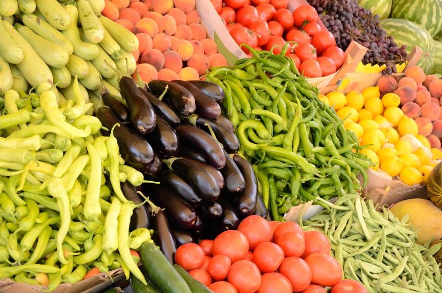
The decay and deterioration of fruits and vegetables during postharvest transportation, storage and marketing is an important factor affecting the quality, shelf life and safety of human consumption. Decay caused by plant pathogenic fungi is the main cause of postharvest losses of fruits and vegetables. Fungi are eukaryotic organisms, and the cell wall is essential for the survival of fungal cells. Degradation of the cell wall can cause the fungal cells to lose their osmotic pressure balance, so destroying the integrity of the cell wall has become a potential antifungal measure. β-1,3-glucan is an important antibacterial protein secreted by biocontrol microorganisms, and plants themselves also induce the production of β-1,3-glucanase during the process of resisting fungal infection [53].
β-1,3-glucanase can degrade the β-1,3-glucan sugar chains in the cell wall, causing the mycelia of pathogenic fungi to break or become deformed, resulting in leakage of the protoplasm of the pathogenic fungi and inhibiting spore germination. In addition, this process can also release fungal cell wall fragment inducers, induce plant immune induction, and indirectly promote the accumulation of phytoalexins in the host plant, increasing its disease resistance [54]. The biological control method based on β-1,3-glucanase can effectively prevent and control fungal diseases in plants. It has the advantages of no drug resistance, targeting only the target pathogen without harming other beneficial organisms, no pesticide residues, and no toxicity or pollution. Therefore, exploring the role of β-1,3-glucanase in postharvest preservation of fruits and vegetables and developing new green biological preservatives based on β-1,3-glucanase is one of the potential development trends in green preservation technology for fruits and vegetables.
Lou Shubao et al. [55] determined the activity of β-1,3-glucanase in soybean leaves and its antibacterial effect on fungi. The results showed that the β-1,3-glucanase activity of the plant reached a peak 48 hours after inoculation with Phytophthora sojae. The antibacterial experiment was carried out using crude β-1,3-glucanase enzyme extract extracted 48 hours after inoculation. It was found that β-1,3 -glucanase crude enzyme solution has a significant inhibitory effect on the mycelial growth and spore germination of Phytophthora sojae. Chen Xiaoyun et al. [56] showed that β-1,3-glucanase has a significant effect on inhibiting the growth of bacteria and disease in fruits such as apples, pears, and bananas after harvest, and can effectively prevent post-harvest rot caused by fungi. This characteristic can be used for the storage and preservation of tropical fruits. Rajninec et al. [57] found that crude β-1,3-glucanase protein from Drosera binate had an inhibitory effect on the growth of Rhizoctonia solani, Alternaria solani and Fusarium poae.
4.2 β-1,3-glucan preparation Functional oligosaccharides
β-1,3-glucan, also known as fucoidan oligosaccharides, is a food functional factor with good biological activity, and has the activity of regulating the body's immunity, resisting infection, and regulating the balance of intestinal flora. Beta-1,3-glucan oligosaccharides prepared by hydrolyzing available polysaccharides or kelp polysaccharides can be used as a new type of prebiotic in functional food development. In addition, some small-molecule soluble beta-1,3-oligosaccharides can be used as immune activators to induce immune responses in plants and thus improve plant disease resistance. In addition, some small-molecule soluble β-1,3-oligosaccharides can be used as immune activators to induce immune responses in plants, thereby improving plant disease resistance. β-1,3-glucanase hydrolyzes β-1,3-glucan to prepare β-1,3-oligosaccharides, which has the advantages of high specificity and few by-products. It is considered to be a promising method for producing oligosaccharides. Wang Yanxin et al. [22] found that the GH55 family β-1,3-glucanase (AcGluA) can hydrolyze kelp polysaccharides into a series of oligosaccharides oligosaccharides, and high doses of oligosaccharides can induce an immune response in rice seedlings, thereby conferring resistance to rice blast. This shows that the hydrolytic products of β-1,3-glucanase have significant biocontrol effects and provide some guidance for the application of β-1,3-oligosaccharides.
Li Kuikui et al. [58] cloned and purified a novel β-1,3-glucanase (GcGluE) from Cellulosimicrobium cellulans. Analysis of substrate specificity and hydrolytic products revealed that the enzyme exhibited the highest hydrolytic activity on renewable polysaccharides, and the main hydrolytic products were disaccharides and trisaccharides. In addition, after the pre-treatment of homogenization, the degradation efficiency of GcGluE on xylan will increase by 7.1 times, which has certain application potential. Gao Minjie et al. [18] obtained β-1,3-glucanase from Trichoderma harzianum, and the enzyme activity could reach up to 198.57 U/mL after 118 h of fermentation. Based on the characteristics of this enzyme, a method for preparing multifunctional oligosaccharides by enzymatic hydrolysis of β-1,3-glucan was established, which makes β-1,3-glucan enzyme has certain application prospects in the industrial production of oligosaccharides.
4.3 Application of β-1,3-glucan in the brewing industry
In addition to its important role in antifungal and oligosaccharide production, β-1,3-glucanase can also be used in the brewing industry. In the brewing industry, barley is the main raw material for beer production. During the production process, some microorganisms secrete high-molecular-weight β-1,3-glucan into the extracellular space, which increases the viscosity of the fermentation liquid, causing difficulties in filtration. Eventually, flocculent gels may form in the beer, reducing beer production and increasing brewing costs. Lü Lili et al. [59] found that if appropriate β-1,3-glucanase is added during the fermentation process, the content of high-molecular-weight glucan can be significantly reduced, thereby reducing the viscosity of the fermentation broth, achieving the purpose of lean beer and improving the filtration process.
4.4 β-1,3-glucan preparation of yeast protoplasts
The main chemical component of the yeast cell wall is β-glucan. One type is the relatively abundant β-1,3-glucan, which forms the skeleton of the yeast cell wall. The other type is the less abundant β-1,6-glucan, which fills the space. The key to preparing yeast protoplasts is to break down the insoluble β-1,3-glucan in the cell wall, so β-1,3-glucanase is an important preparation for protoplasts [60]. Duan Huike et al. [61] used the β-1,3-glucanase produced by the Trichoderma strain LE02 to enzymolyze and solubilize the beer yeast glucan. Macromolecular water-soluble yeast glucan can also be obtained by β-1,3-glucanase enzymolysis technology and ultrafiltration separation technology.
4.5 β-1,3-glucan removes biological films
Biofilms are complex structures composed of microorganisms and their extracellular secretions. Key spoilage bacteria in food such as Salmonella and Pseudomonas aeruginosa produce biofilms, which can reduce the bactericidal effect of commonly used disinfectants and antibiotics, posing a food safety risk. β-1,3-glucan is an important component of the biofilm of Candida albicans and plays an important role in the extracellular matrix of the biofilm. In contrast, β-1,3-glucanase has a certain effect on removing the biofilm and can control spoilage bacteria in the food industry. Nett et al. [62] found that treating Candida with low concentrations of β-1,3-glucanase significantly enhanced the effect of the antifungal drugs fluconazole and amphotericin B on the fungus. Mitchell et al. [63] also demonstrated experimentally that the sensitivity of the antifungal drug gradually increased with the hydrolysis of β-1,3 glucan in the extracellular matrix, which indicates that β-1,3-glucanase has a certain effect in removing biological films.
4.6 Synergistic effect of β-1,3-glucan and chitinase
Both β-1,3-glucanase and chitinase have the effect of degrading β-1,3-glucan and chitin as well as peptidoglycan in the fungal cell wall. Both enzymes have broad-spectrum resistance in the defense against plant pests and diseases, which can reduce the use of chemical pesticides and lower environmental pollution. Mauch et al. [64] found that the combined action of chitinase and β-1,3-glucanase has a better antibacterial effect than the individual enzymes, indicating that the two enzymes have a synergistic effect in inhibiting the growth of pathogenic bacteria. Cota et al. [65] found that the synergistic effect of β-1,3-glucanase and chitinase can significantly resist the impact of Alternaria alternata on stored tomatoes. This shows that β-1,3-glucanase and chitinase have a synergistic effect on the post-harvest antibacterial preservation of fruits and vegetables, and have a stronger antibacterial ability than a single strain, so they have good application prospects.
5 Conclusion
Because β-1,3-glucan can specifically hydrolyze β-1,3-glucan, it has important application prospects in the fields of functional oligosaccharide preparation, fruit and vegetable preservation, biomedicine, and plant disease resistance. Researchers have already studied the structure, function and application of a series of different family β-1,3-glucanases. In this context, the focus of subsequent research on β-1,3-glucanases will be on how to obtain new β-1,3-glucanases with good application properties and achieve their efficient fermentation preparation.
However, due to the complexity of natural β-1,3-glucan substrates, the catalytic efficiency of existing β-1,3-glucanases for different types of substrates still needs to be further improved, and enzymatic hydrolysis of some insoluble β-1,3-glucan substrates is still difficult. In addition, research on enzyme structure and function is an important basis for exploring the catalytic mechanism of enzymes, mining the catalytic properties of enzymes, and conducting research on enzyme molecular modification. In view of the complexity of natural β-1,3-glucan substrates and the diversity of β-1,3-glucanase families, it is a development trend to study the differences in substrate binding and catalytic mechanisms of different β-1,3-glucanase families, clarify the substrate recognition mechanism of β-1,3-glucanase for complex glucan molecules, and explore a multi-enzyme combination catalytic system based on β-1,3-glucanase in order to achieve the efficient application of β-1,3-glucanase. 1,3-glucanase is the development trend for its efficient application.
Reference:
[1] KADAM S U, TIWARI B K, O’DONNELL C P. Extraction, structure and biofunctional activities of laminarin from brown algae[J] . International Journal of Food Science and Technology, 2015, 50(1): 24-31. DOI:10.1111/ijfs.12692.
[2] MIYOSHI K, UEZU K, SAKURAI K, et al. Proposal of a new hydrogen-bonding form to maintain curdlan triple helix[J]. Chemistry & Biodiversity, 2004, 1(6): 916-924. DOI:10.1002/cbdv.200490073.
[3] Fu Yunbin, Zhao Xiaoming, Du Yuguang. Research progress on the biological activity and application of cydramon polysaccharide and its derivatives [J]. Food Science, 2012, 33(7): 315-319.
[4] SHIMIZU J, TSUCHIHASHI N, KUDOH K, et al. Dietary curdlan increases proliferation of bifidobacteria in the cecum of rats[J]. Bioscience Biotechnology and Biochemistry, 2001, 65(2): 466-469. DOI:10.1271/bbb.65.466.
[5] Yu Yonghua, Xu Feifei, Lin Lin, et al. Research progress on the hypolipidemic effect of oat β-glucan [J]. Journal of Clinical and Pathological Sciences, 2022, 42(2): 486-491. DOI:10.3978 / j.issn.2095-6959.2022.02.034.
[6] DAVIES G, HENRISSAT B. Structures and mechanisms of glycosyl hydrolases[J]. Structure, 1995, 3(9): 853-859. DOI:10.1016/S0969- 2126(01)00220-9.
[7] ILARI A, FIORILLO A, ANGELACCIO S, et al. Crystal structure of a Famil16 endoglucanase from the hyperthermophile basis of substrate recognition[J]. FEBS Journal, 2009, 276(4): 1048-1058. DOI:10.1111/ j.1742-4658.2008.06848.x.
[8]YUAN Y, ZHANG X, ZHANG H, et al. Degradative GH5 β-1,3- 1,4-glucanase PpBglu5A for glucan in Paenibacillus polymyxa KF-1[J]. Process Biochemistry, 2020, 98: 183-192. DOI:10.1016/ j.procbio.2020.08.008.
[9]JAAFAR N R, KHOIRI N M, ISMAIL N F, et al. Functional characterisation and product specificity of endo-β-1,3-glucanase from alkalophilic bacterium, Bacillus lehensis G1[J] . Enzyme and Microbial Technology, 2020, 140: 109625.
[10] SHI P, YAO G, YANG P, et al. Cloning, characterization, and antifungal activity of an endo-1,3-β-D-glucanase from Streptomyces sp. S27[J]. Applied Microbiology and Biotechnology, 2009, 85(5): 1483-1490. DOI:10.1007/s00253-009-2187-1.
[11]HARTL L, GASTEBOIS A,AIMANIANDAV, et al. Characterization of the GPI-anchored endo β-1,3-glucanase Eng2 of Aspergillus fumigatus[J]. Fungal Genetics and Biology, 2011, 48(2): 185-191. DOI:10.1016/j.fgb.2010.06.011.
[12]DA SILVA AIRES R, STEINDORFF A S, RAMADA M H S, et al. Biochemical characterization of a 27 kDa 1,3-β-D-glucanase from Trichodermaasperellum induced by cell wall of Rhizoctonia solani[J].Carbohydrate Polymers, 2012, 87(2): 1219-1223. DOI:10.1016/ j.carbpol.2011.09.001.
[13] ISHIDA T, FUSHINOBU S, KAWAI R, et al. Crystal structure of glycoside hydrolase family 55β-1,3-glucanase from the basidiomycete Phanerochaetechrysosporium[J]. Journal of Biological Chemistry, 2009, 284(15): 10100-10109.
[14] VARGHESE J N, GARRETT T P, COLMAN P M, et al. Three- dimensional structures of two plant β-glucan endohydrolases with distinct substrate specificities[J]. Proceedings of the National Academy of Sciences of the United States of America, 1994, 91(7): 2785-2789.
[15]ROMERO I, FERNANDEZ-CABALLERO C, GONI O, et al. Functionality of a class I β-1,3-glucanase from skin of table grapes berries[J] . Plant Science, 2008, 174(6): 641-648. DOI:10.1016 / j.plantsci.2008.03.019.
[16] YU W Q, ZHENG G P, QIU D W, et al. Paenibacillus terrae NK3-4: a potential biocontrol agent that produces β-1,3-glucanase[J]. Biological Control, 2019, 129: 92-101. DOI:10.1016/j.biocontrol.2018.09.019.
[17]LI C, WEN Y, HE Y, et al. Purification and characterization of a novel β-1,3-glucanase from Arca inflata and its immune- enhancing effects[J]. Food Chemistry, 2019, 290: 1-9. DOI:10.1016/ j.foodchem.2019.03.131.
[18]GAO Minjie, YAN Jiajun, ZHAO Yue, et al. Expression of a thermostable β-1,3-glucanasefrom Trichoderma harzianum in Pichia pastoris and use in oligoglucosides hydrolysis[J] . Process Biochemistry, 2021, 107: 74-82.
[19]ZHANG D, SPADARO D, VALENTE S, et al. Cloning, characterization and expression of an exo-1,3-β-glucanase gene from the antagonistic yeast, Pichia guilliermondii strain M8 against grey mold on apples[J]. Biological Control, 2011, 59(2): 284-293. DOI:10.1016/j.biocontrol.2011.06.018.
[20]KALYANI D C, REICHENBACH T, ASPEBORG H, et al. A homodimeric bacterial exo-β-1,3-glucanase derived from moose rumen microbiome shows a structural framework similar to yeast exo-β-1,3- glucanases[J]. Enzyme and Microbial Technology, 2021, 143: 109723. DOI:10.1016/j.enzmictec.2020.109723.
[21]YI P, YAN Q, JIANG Z, et al. A first glycoside hydrolase family 50 endo-β-1,3-D-glucanase from Pseudomonas aeruginosa[J] . Enzyme and Microbial Technology, 2018, 108: 34-41. DOI:10.1016/ j.enzmictec.2017.09.002.
[22]WANG Yanxin, ZHAO Yuqiang, WANG Xiaowen, et al. Functional characterization of the novel laminaripentaose-producing β-1,3- glucanase MoGluB and its biocontrol of Magnaporthe oryzae[J]. Journal of Agricultural and Food Chemistry, 2021, 69(33): 9571-9584.
[23] Cheng Zhengxiang. Expression of β-1,3-glucanase in Pichia pastoris [D]. Harbin: Harbin Institute of Technology, 2013: 5-10.
[24] FENG Jianwei, XU Shenyuan, FENG Ruirui, et al. Identification and structural analysis of a thermophilic β-1,3-glucanase from compost[J]. Bioresources and Bioprocessing, 2021, 8(1): 102-112. DOI:10.1186/ s40643-021-00449-4.
[25]MUHAMMED M T, SON Ç D, İZGÜ F. Three dimensional structure prediction of panomycocin, a novel exo-β-1,3-glucanase isolated from Wickerhamomyces anomalus NCYC 434 and the computational site-directed mutagenesis studies to enhance its thermal stability for therapeutic applications[J]. Computational Biology and Chemistry, 2019, 80: 270-2777. DOI:10.1016/j.compbiolchem.2019.04.006.
[26]FIBRIANSAH G, MASUDA S, KOIZUMI N, et al. The 1.3 angstrom crystal structure of a novel endo-β-1,3-glucanase of glycoside hydrolase family 16 from alkaliphilic Nocardiopsis sp. strain F96[J]. Proteins, 2007, 69(3): 683-690.
[27]VUONG T V, WILSON D B. Glycoside hydrolases: catalytic base/nucleophile diversity[J]. Biotechnology and Bioengineering, 2010, 107(2): 195-205. DOI:10.1002/bit.22838.
[28]ILARI A, FIORILLO A, ANGELACCIO S, et al. Crystal structure of a family 16 endoglucanase from the hyperthermophile Pyrococcus furiosus-structural basis of substrate recognition[J]. The FEBS Journal, 2009, 276(4): 1048-1058. DOI:10.1111/j.1742-4658.2008.06848.x.
[29]ODA M, INABA S, KAMIYAN, et al. Structural and thermodynamic characterization of endo-1,3-β-glucanase: insights into the substrate recognition mechanism[J]. BBA-Proteins Proteomics, 2018, 1866(3): 415-425.
[30]JENG W Y, WANG N C, LIN C T, et al. Crystal structures of the laminarinase catalytic domain from Thermotoga maritima MSB8 in complex with inhibitors essential residues for β-1,3-and β-1,4-glucan selection[J]. Journal of Biological Chemistry, 2011, 286(52): 45030-45040.
[31]HONG T Y, HSIAO YY, MENG M, et al. The 1.5 angstrom structure of endo-1,3-β-glucanase from Streptomyces sioyaensis: evolution of the active-site structure for 1,3-β-glucan-binding specificity and hydrolysis[J]. Acta Crystallographica Section D: Structural Biology, 2008, 64: 964-970. DOI:10.1107/s0907444908021550.
[32]BLEICHER L, PRATES E T, GOMES T C F, et al. Molecular basis of the thermostability and thermophilicity of laminarinases: X-ray structure of the hyperthermostable laminarinase from Rhodothermus marinus and molecular dynamics simulations[J]. Journal of Physical Chemistry B, 2011, 115(24): 7940-7949. DOI:10.1021/jp200330z.
[33]LABOUREL A, JAM M, JEUDY A, et al. The β-glucanaseZgLamA from Zobellia galactanivorans evolved a bent active site adapted for efficient degradation of algal laminarin[J]. Journal of Biological Chemistry, 2014, 289(4): 2027-2042.
[34]YANG J, XU Y Q, MIYAKAWA T, et al. Molecular basis for substrate recognition and catalysis by a marine bacterial laminarinase[J]. Applied and Environmental Microbiology, 2020, 86(23): 15. DOI:10.1128/aem.01796-20.
[35]WOJTKOWIAK A, WITEK K, HENNIG J, et al. Structures of an active-site mutant of a plant 1,3-β-glucanase in complex witholigosaccharide products of hydrolysis[J]. Acta Crystallographica Section D: Biological Crystallography, 2013, 69: 52-62. DOI:10.1107/s0907444912042175.
[36]RECEVEUR-BRECHO T V, CZJ ZEK M , BARRE A , et al. Crystal structure at 1.45-Å resolution of the major allergen endo- β-1,3-glucanase of banana as a molecular basis for the latex-fruit syndrome[J]. Proteins, 2006, 63(1): 235-242.
[37]UNFRIED F, BECKER S, ROBB C S, et al. Adaptive mechanisms that provide competitive advantages to marine bacteroidetes during microalgal blooms[J] . ISME Journal, 2018, 12(12): 2894-2906. DOI:10.1038/s41396-018-0243-5.
[38]TAKASHIMA T, TAKU T, YAMANAKA T, et al. Crystal structure and biochemical characterization of CJP38, a β-1,3-glucanase and allergen of Cryptomeria japonica pollen[J]. Molecular Immunology, 2019, 116: 199-207.
[39]WU H M, LIU S W, HSU M T, et al. Structure, mechanistic action, and essential residues of a GH-64 enzyme, laminaripentaose-producing β-1,3-glucanase[J]. Journal of Biological Chemistry, 2009, 284(39): 26708-26715.
[40]QIN Zhen, YANG Dong, YOU Xin, et al. The recognition mechanism of triple-helical β-1,3-glucan by a β-1,3-glucanase[J]. Chemical Communications, 2017, 53(67): 9368-9371. DOI:10.1039/c7cc03330c.
[41] GHOSH R, CHAKRABARTI C. Crystal structure analysis of NP24-I: a thaumatin-like protein[J]. Planta, 2008, 228(5): 883-890. DOI:10.1007/s00425-008-0790-5.
[42]HENRISSAT B, GARRON M L . How a glycoside hydrolase recognizes a helical polyglucan[J]. Structure, 2017, 25(9): 1319-1321. DOI:10.1016/j.str.2017.08.004.
[43]KUMAR K, CORREIA M A S, PIRES V M R, et al. Novel insights into the degradation of β-1,3-glucansby the cellulosome of Clostridium thermocellum revealed by structure and function studies of a family 81 glycoside hydrolase[J]. International Journal of Biological Macromolecules, 2018, 117: 890-901. DOI:10.1016/ j.ijbiomac.2018.06.003.
[44] ZHOU Peng, CHEN Zhongzhou, YAN Qiaojuan, et al. The structure of a glycoside hydrolase family 81 endo-β-1,3-glucanase[J]. Acta Crystallographica Section D: Structural Biology, 2013, 69: 2027-2038. DOI:10.1107/s090744491301799x.
[45]MA Junwen, QIN Zhen, ZHOU Peng, et al. Structural insights into the substrate recognition and catalytic mechanism of a fungal glycoside hydrolase family 81 β-1,3-glucanase[J]. Enzyme and Microbial Technology, 2022, 153: 109948. [46] PLUVINAGE B, FILLO A, MASSEL P, et al. Structural analysis of a family 81 glycoside hydrolase implicates its recognition of β-1,3- glucan quaternary structure[J]. Structure, 2017, 25(9): 1348-1359. DOI:10.1016/j.str.2017.06.019.
[47] BIANCHETTI C M, TAKASUKAT E, DEUTSCH S, et al. Active site and laminarin binding in glycoside hydrolase family 55[J]. Journal of Biological Chemistry, 2015, 290(19): 11819-11832. DOI:10.1074/jbc. M114.623579.
[48] PAPAGEORGIOU A C, CHEN J, LI D. Crystal structure and biological implications of a glycoside hydrolase family 55 β-1,3- glucanase from Chaetomium thermophilum[J] . Biochimica et Biophysica Acta-Proteins and Proteomics, 2017, 1865(8): 1030-1038. DOI:10.1016/j.bbapap.2017.05.002.
[49] JIA X, WANG C, DU X, et al. Specific hydrolysis of curdlan with a novel glycoside hydrolase family 128 β-1,3-endoglucanase containing a carbohydrate-binding module[J]. Carbohydrate Polymers, 2021, 253: 117276-117286.
[50] SANTOS C R, COSTA P, VIEIRA P S, et al. Structural insights into β-1,3-glucan cleavage by a glycoside hydrolase family[J]. Nature Chemical Biology, 2020, 16(8): 920-929. DOI:10.1038/s41589-020- 0554-5.
[51] GASTEBOIS A, AIMANIANDA V, BACHELLIER-BASSI S, et al. SUN proteins belong to a novel family of β-(1,3)-glucan-modifying enzymes involved in fungal morphogenesis[J]. Journal of Biological Chemistry, 2013, 288(19): 13387-13396.
[52] DÉJEAN G, TAMURA K, CABRERA A, et al. Synergy between cell surface glycosidases and glycan-binding proteins dictates the utilization of specific β(1,3)-glucans by human gut Bacteroides[J]. mBio, 2020, 11(2): 1-21.
[53]YAN F, YE X L, LI C H, et al. Isolation, purification, gene cloning and expression of antifungal protein from Bacillus amyloliquefaciens MG - 3 [ J] . Food Chemistry, 2 0 2 1 , 3 4 9 : 7 . DOI : 1 0 . 1 0 1 6 / j.foodchem.2021.129130.
[54] FESEL P H, ZUCCARO A.β-Glucan: crucial component of the fungal cell wall and elusive MAMPin plants[J]. Fungal Genetics and Biology, 2016, 90: 53-60. DOI:10.1016/j.fgb.2015.12.004.
[55] Lou Shubao, Peng Dongjun, Wang Hui. Bacteriostatic activity of soybean β-1,3-glucanase [J]. Journal of Heilongjiang Bayi Agricultural University, 2008 (3): 27-29.
[56] Chen Xiaoyun, Li Jianbin, Lin Ying, et al. Application of β-1,3-glucanase and chitinase in the preservation of tropical fruits [J]. Food Industry Science and Technology, 2008(5): 294-296.
[57]RAJNINEC M, FRATRIKOVA M, BOSZORADOVA E, et al. Basic β-1,3-glucanase from Drosera binata exhibits antifungal potential in transgenic tobacco plants[J] . Plants, 2021, 10(8): 1747-1762. DOI:10.3390/plants10081747.
[58]LI Kuikui, CHEN Wei, WANG Wenxia, et al. Effective degradation of curdlan powder by a novel endo-β-1,3-glucanase[J]. Carbohydrate Polymers, 2018, 201: 122-130. DOI:10.1016/j.carbpol.2018.08.048.
[59] Lv Lili, Wang Ruibin, Wang Jialin, et al. Effect of high-efficiency β-glucanase on the filtration rate of wort [J]. Brewing Science and Technology, 2010(3): 75-77. DOI:10.13746/j.njkj.2010.03.028.
[60] Lin Kaijiang, Ruan Lijuan, Wang Longying. Research on the preparation of yeast protoplasts with cellulase [J]. Science and Technology Bulletin, 1996, 12(2): 118-121.
[61] Duan Huike, Xiong Shanbo, Liu Haimei. Enzymatic solubilization of yeast β-1,3-glucan and product analysis [J]. Food Science, 2008, 29(1): 185-189.
[62] NETT J, LINCOLN L, MARCHILLO K, et al. Putative role of β-1,3 glucans in Candida albicans biofilm resistance[J]. Antimicrobial Agents and Chemotherapy, 2007, 51(2): 510-520. DOI:10.1128/ aac.01056-06.
[63]MITCHELL K F, TAFF H T, CUEVAS MA, et al. Role of matrix β-1,3 glucan in antifungal resistance of non-albicans Candida Biofilms[J]. Antimicrobial Agents and Chemotherapy, 2013, 57(4): 1918-1920. DOI:10.1128/aac.02378-12.
[64] MAUCH F, MAUCH-MANI B, BOLLER T. Antifungal hydrolases in pea tissue: II. inhibition of fungal growth by combinations of chitinase and β-1,3-glucanase[J] . Plant Physiology, 1988, 88(3): 936-942.
[65]COTA I E, TRONCOSO -ROJAS R, SOTELO -MUNDO R, et al. Chitinase and β-1,3-glucanase enzymatic activities in response to infection by Alternaria alternata evaluated in two stages of development in different tomato fruit varieties[J] . Scientia Horticulturae, 2007, 112(1): 42-50 .


