

 English
 French
 Spanish
 Russian
 Korean
 Japanese
Study on Lutein and Zeaxanthin for Eye Health
Zeaxanthin and lutein play an important role in protecting visual function, fighting cancer, preventing the occurrence and development of cardiovascular diseases, and improving the body's immune system. They are the main pigment components that make up the macular region of the human retina, and can filter out a large amount of harmful blue light, exerting anti-inflammatory and antioxidant effects [1]. Zeaxanthin and lutein have been widely used in many fields and have achieved certain results in the prevention and treatment of ophthalmic diseases. In this article, the authors provide a review of the research progress of zeaxanthin and lutein in ophthalmology in recent years.
1 Properties and sources of zeaxanthin and lutein
The molecular formulas of zeaxanthin and lutein are both C40 H56 O2. They are isomers of each other and are carotenoids that are widely found in nature. To date, more than 600 carotenoids have been discovered by humans, among which 30 to 50 types have been proven to be able to be replenished by the human body through the diet in a timely manner [2]. The molecular structure of zeaxanthin and lutein contains single bonds and double bonds that are distributed alternately. The presence of conjugated double bonds gives them peroxyl radicals, which can effectively quench the transfer of oxygen free radicals, thereby exerting an antioxidant effect. Zeaxanthin and lutein are widely found in dark green vegetables. They cannot be synthesized directly by the human body and must be obtained through dietary or other means, such as eating spinach, kale, pumpkin and zucchini, etc. [3].

2 Distribution of zeaxanthin and lutein
Zeaxanthin and lutein are found in all tissues of the body, with the most prominent being the eye tissue, and together with the total amount contained in brain tissue, they account for 80% to 90% of the entire body [4]. Zeaxanthin is mainly found in the fovea of the retina, while lutein is mainly found in the surrounding area of the retina [5]. The concentration of zeaxanthin in the fovea is twice that of lutein. As the distance from the fovea increases, zeaxanthin is gradually replaced by lutein, becoming the main carotenoid in the peripheral retina [5]. The macular pigment is composed of zeaxanthin and lutein, which can effectively quench oxygen free radicals and reduce blue light damage, thereby protecting eye tissue from irritation and damage [6]. A decrease in macular pigment optical density (MPOD) will lead to irreversible structural damage and a decrease in the function of the retina, thereby causing the occurrence and development of retinal diseases [6].
3 Research progress of zeaxanthin and lutein in ophthalmic diseases
3.1 Retinopathy of prematurity (ROP)
ROP is one of the main causes of blindness in children and is closely related to extremely low birth weight and a history of oxygen inhalation [7]. Gong et al. [8] found using a meta-analysis that intervention with zeaxanthin and lutein can significantly reduce the risk of ROP in premature infants. A study of 150 newborns showed that timely supplementation with lutein after birth can improve the body's antioxidant capacity and and reduced hydrogen peroxide levels in the body [9]. Manzoni et al. [10] compared breast milk and formula-fed extremely low birth weight infants through analysis, and the results showed that the risk of breast milk-fed extremely low birth weight infants suffering from ROP was significantly lower than that of formula-fed infants, which may be related to the fact that breast milk is rich in zeaxanthin and lutein.
3.2 Retinitis pigmentosa (RP)
RP is a hereditary retinal degenerative disease in which genetic defects play a decisive role. It is manifested by apoptosis of photoreceptor cells, damage to retinal function and structure, abnormalities and damage to the retinal pigment epithelium, and ultimately complete loss of central vision. Researchers are still divided on whether carotenoids can improve RP. Bahrami et al. [11] compared RP patients in the experimental group who took lutein supplements with those in the control group who were given a placebo. The results showed that the visual fields of the experimental group of RP patients improved significantly and were statistically significant. In contrast, Aleman et al. [12] reported that although the lutein concentration in the serum of RP patients increased after oral lutein supplementation, there was no significant improvement in the patients' central visual acuity. The difference in this result may be due to the different dosages and periods of lutein administration to RP patients.
3.3 Diabetic retinopathy (DR)
DR is one of the common causes of blindness. The development of DR is closely related to the course of diabetes and the degree of control. The formation of new blood vessels in the retina is the main manifestation of DR, and abnormal and disordered glucose metabolism is the root cause of DR. Research reports have shown that DR is closely related to the degree of oxidative damage in the body, and the degree of oxidative damage in DR patients is significantly higher than in people with diabetes who have not yet developed DR [13]. Tang et al. [14] used DR model mice as the research object, and the results showed that the experimental group supplemented with zeaxanthin and lutein achieved some degree of improvement in retinopathy compared to the control group that was not given supplements. Gong et al. [8] found that increasing the serum concentrations of zeaxanthin and lutein can significantly reduce the damage caused by oxidative stress and has a positive preventive effect on the development of DR. Therefore, zeaxanthin and lutein can be used as one of the effective adjuvant means to prevent and treat DR in diabetic patients.
3.4 Glaucoma
Glaucoma is a disease characterized by characteristic optic nerve atrophy, visual field defects, and progressive retinal ganglion cell (RGC) damage. RGC) damage is the main feature of this disease. It is one of the main causes of blindness and is characterized by increased pathological intraocular pressure. Zhang et al. [15] used a rabbit model with RGC damage as the research object and found that lutein can reduce retinal toxicity by regulating the mitogen-activated protein kinase signaling pathway, further increasing the number of RGCs, thereby providing necessary protection for the optic nerve.
Coleman et al. [16] studied 1,155 women over the age of 65 and found that eating green vegetables and fruits rich in vitamins A, C and beta-carotene can significantly reduce the risk of glaucoma. Subsequently, Giaconi et al. [17] also confirmed this conclusion, and further pointed out that eating fresh oranges, peaches, carrots, spinach and kale can effectively reduce the risk of glaucoma, and is statistically significant. Ramdas et al. [18] followed up 3,502 cases of glaucoma patients over the age of 55. The results showed that taking carotenoid supplements and vitamin B1 can significantly reduce the risk of further development of open-angle glaucoma, while a high intake of magnesium-rich foods will greatly increase the risk of open-angle glaucoma, and it is three times higher in people with low magnesium intake.
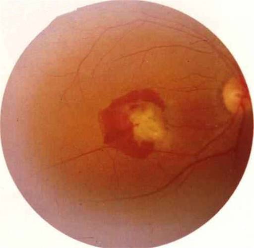
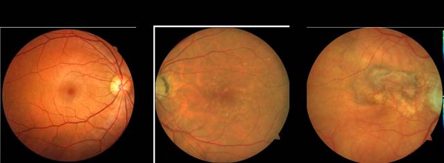
3.5 Age-related macular degeneration (AMD)
AMD is one of the factors that cause severe irreversible blindness in middle-aged and elderly people [19]. The pathogenesis of AMD is still unclear, but it is mainly believed to be closely related to factors such as age, oxidative stress damage, blue light damage, genetic factors, environmental factors, inflammation, and autoimmune diseases [20]. AMD has made breakthroughs through the injection of anti-vascular endothelial growth factor (VEGF), which has significantly improved clinical symptoms. However, there are many problems, such as the high price of anti-VEGF injections and complications caused by repeated injections [21]. Studies have shown that the concentration of zeaxanthin and lutein in the blood directly affects the MPOD level, and the risk of AMD in people with low MPOD is significantly higher [22].
Studies have shown that zeaxanthin and lutein can effectively reduce the risk of AMD, delay the progression of AMD, and have a certain therapeutic effect on AMD [23-24]. Ma et al. [25] used a meta-analysis to explore the relationship between AMD and zeaxanthin and lutein. The results showed that intake of zeaxanthin and lutein supplements can significantly reduce the risk of early AMD by 4%, and for reducing the risk of late AMD, the rate can be as high as 26%. Chew et al. [26] followed up on AMD patients for 10 years, the results showed that zeaxanthin and lutein can effectively delay the progression of AMD. Weigert et al. [27] studied 126 AMD patients over the age of 50 and found that taking zeaxanthin and lutein supplements can increase the retinal MPOD of AMD patients, thereby significantly improving visual acuity.
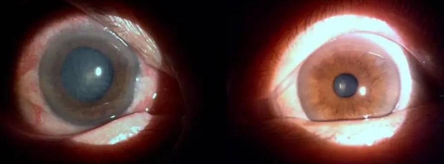
3.6 Age-related cataract (ARC)
ARC, also known as senile cataract, is most common in people over 50 years old. It can cause the lens of the middle-aged and elderly to become cloudy, and the occurrence of the disease is closely related to age. The pathogenesis of ARC is not yet clear, but it is mainly believed to be related to oxidative stress damage, genetics, and environmental factors. Among these, oxidative stress damage is one of the main causes of ARC, while antioxidants can protect the lens and reduce the damage caused by oxidative stress [28].
Gao et al. [29] found in a study of a model of oxidative damage to lens epithelial cells that intervention with zeaxanthin and lutein significantly reduced the degree of oxidative damage to lens epithelial cells, protected lens proteins and lipids, and that lutein and zeaxanthin supplementation can effectively reduce the risk of ARC. Liu et al. [30] compared groups with higher and lower serum lutein and zeaxanthin concentrations through a meta-analysis, and the results showed that the higher concentration group could significantly reduce the risk of nuclear cataracts, and the difference was statistically significant. For cortical cataracts and posterior subcapsular cataracts, although the risk of developing these cataracts could be reduced, the difference was not statistically significant. Ma et al. [31] also confirmed the above conclusions, and further pointed out that increasing the intake of zeaxanthin and lutein can effectively reduce the risk of nuclear cataracts by 25%, posterior subcapsular cataracts by 23%, and cortical cataracts by 15%.
4 Safety assessment of zeaxanthin and lutein
Shao et al. [32] conducted a risk assessment of lutein supplements and found that the safe intake of lutein is 20 mg·d-1. Subsequently, 4,203 AMD patients who took lutein and zeaxanthin supplements were followed up for 5 years and found that, apart from some patients experiencing yellowing of the skin, no other side effects occurred [33]. However, a recent report indicated that a woman who had taken lutein supplements at a dose of 20 mg·d−1 for 8 years experienced lens dissolution in her right eye and no abnormalities in her left eye after ceasing intake for 7 months [34]. Therefore, the safe dose range for long-term intake of zeaxanthin and lutein supplements needs to be further explored and studied.

5 Summary and outlook
Zeaxanthin and lutein play an extremely important role in vision protection, disease prevention, boosting the body's immune system and maintaining good health. In recent years, with the continuous exploration and research on zeaxanthin and lutein, many breakthroughs have been made in the prevention and treatment of eye diseases such as AMD, ARC, DR and glaucoma. However, there are still many problems that need to be further addressed, such as the mechanism of zeaxanthin and lutein in preventing and treating diseases has not been fully elucidated, the therapeutic dose of zeaxanthin and lutein given to patients and the duration of the course of treatment have not been standardized, and further research is needed to determine whether there are toxic reactions associated with long-term high intake of zeaxanthin and lutein.
Reference:
[1]PHELAN D,PRADO-CABRERO A,NOLAN J M.Analysis of lutein,zeaxanthin,and meso-zeaxanthin in the organs of carotenoid-supplemented chickens[J].Foods,2018,7(2) : 1-8.
[2]BERNSTEIN P S.The role of lutein and zeaxanthin in protection against age-related macular degeneration[J].Acta Hor- tic,2016,1106( 1106) : 153-160.
[3]THIRUPATHI B,RAO G S,SWAPNA V,et al.HPLC analysis of lutein and zeaxanthin in green and colored varieties of vege- tables[J].International Journal of Pharmacognosy and Phyto- chemical Research,2017,9(7) : 11008-11010.
[4]VISHWANATHAN R , SCHALCH W,JOHNSON E J.Macular pigment carotenoids in the retina and occipital cortex are relat- ed in humans[J].Nutr Neurosci,2015,19(3) : 95-101 .
[5]BERNSTEIN P S,LI B,VACHALI P P,et al.Lutein,zeaxan- thin,and meso-zeaxanthin : the basic and clinical science underlying carotenoid-based nutritional interventions against ocular disease[J].Prog Retin Eye Res,2015,50: 34-66 .
[6]WESTRUP V M Z,DIETZEL M,ZEIMER M,et al.Changes of macular pigment optical density in elderly eyes : a longitudinal analysis from the MARS study[J].Int J Retin Vitr,2016,2 ( 1) : 1-8.
[7] Xue Juxia, Wang Junling, Liu Yao. Nursing experience in the screening of retinopathy of prematurity [J]. Modern Medicine, 2006, 34(2): 139.
[8]GONG X,RUBIN L P. Role of macularxanthophylls in preven- tion of common neovascular retinopathies : retinopathy of pre- maturity and diabetic retinopathy[J].Arch Biochem Biophys, 2015,572( 1) :40-48 .
[9]PERRONE S,TEI M,LONGINI M,et al.Lipid and protein oxi- dation in newborn infants after lutein administration[J].Oxid Med Cell Longev,2014,2014(6) : 1-7.
[10] MANZONI P,STOLFI I,PEDICINO R , et al.Human milk feeding prevents retinopathy of prematurity ( ROP) in pre- term VLBW neonates[J].Early Hum Dev,2013,1 ( 2 ) :64-68.
[11] BAHRAMI H,MELIA M,DAGNELIE G.Lutein supplemen- tation in retinitis pigmentosa : PC-based vision assessment in a randomized double-masked placebo-controlled clinical trial [J].Bmc Ophthalmology,2006,6( 1) : 1-12.
[12] ALEMAN T S,DUNCAN J L,BIEBER M L,et al.Macular pigment and lutein supplementation in retinitis pigmentosa and Usher syndrome[J].Invest Ophthalmol Vis Sci,2001,42 (8) : 1873-1881.
[13]KUNDU D,MANDAL T,NANDI M,et al.Oxidative stress in diabetic patients with retinopathy[J].Ann Afr Med,2014,13 ( 1) :41-46 .
[14]TANG L,ZHANG Y,JIANG Y,et al.Dietary wolfberry amel- iorates retinal structure abnormalities in db / db mice at the early stage of diabetes[J].Exp Biol Med,2011,236 (9 ) :1051-1063.
[15]ZHANG C,WANG Z,ZHAO J,et al.Neuroprotective effect of lutein on NMDA-induced retinal ganglion cell injury in rat retina[J].Cell Mol Neurobiol,2016,36(4) : 531-540 .
[16]COLEMAN A,STONE K G,YU F,et al.Glaucoma risk and the consumption of fruits and vegetables among older women in the study of osteoporotic fractures[J].Am J Ophthalmol, 2008,145 (6) : 1081-1089.
[17] GIACONI J A,YU F,STONE K L,et al.The association of consumption of fruits / vegetables with decreased risk of glau- coma among older African-American women in the study of osteoporotic fractures[J].Am J Ophthalmol,2012,154 (4) :635-644.
[18]RAMDAS W D,WOLFS R C W,JONG K D,et al.Nutrient intake and risk of open-angle glaucoma : the Rotterdam Study [J].Eur J Epidemiol,2012,27(5) : 385-393 .
[19] Wang Weiqiang, Xi Tao, Shen Zilong, et al. Effects of the nitric oxide regulator ZX-5 on the expression of eNOS and ERK proteins in vascular endothelial cells [J]. Journal of Southeast University (Medical Sciences), 2007, 26(6): 417-420.
[20]MENNO V L C,JENNIFER L,YASPAN B L,et al.Mecha- nisms of age-related macular degeneration and therapeutic opportunities[J].J Pathol,2014,232(2) : 151-164.
[21]Cao L, Luan J. Evaluation of the efficacy of intravitreal injection for macular edema secondary to retinal vein occlusion [J]. Journal of Southeast University (Medical Sciences), 2015, 34(1): 147-151.
[22]MA L,LIU R , DUJ H,et al.Lutein,zeaxanthin and meso-zea- xanthin supplementation associated with macular pigment op- tical density[J].Nutrients,2016,8(7) :426-439 .
[23]SCRIPSEMA N K,HU D N,ROSEN R B.Lutein,zeaxanthin, and meso-zeaxanthin in the clinical management of eye dis- ease[J].J Ophthalmol,2015,2015 (6) : 1-13.
[24]PINAZODURN M D,G6MEZULLA F,ARIAS L,et al.Do nutritional supplements have a role in age macular degenera- tion prevention? [J].J Ophthalmol,2014,2014( 1) : 1-15.
[25]MA L,DOU H L,WU Y Q,et al.Lutein and zeaxanthin in- take and the risk of age-related macular degeneration : a sys- tematic review and meta-analysis[J].Brit J Nutr,2012,107 (3) : 350-359 .
[26]CHEW E Y,CLEMONS T E,AGR6N E,et al.Ten-year follow-up of age-related macular degeneration in the age-related eye disease study : AREDS report no.36[J].Jama Ophthal- mol,2014,132(3) : 272-277 .
[27]WEIGERT G,KAYA S,PEMP B,et al.Effects of lutein supplementation on macular pigment optical density and visual acuity in patients with age-related macular degeneration[J].
Invest Ophthalmol Vis Sci,2011,52( 11) : 8174-8178.
[28]ZHENG J S,RAUTIAINEN S,LINDBLAD B E,et al.High- dose supplements of vitamins C and E,low-dose multivitamins,and the risk of age-related cataract: a population-based prospective cohort study of men[J].Am J Epidemiol,2013, 177(6) : 548-555 .
[29] GAO S,QIN T,LIU Z,et al.Lutein and zeaxanthin supple- mentation reduces H2 O2 -induced oxidative damage in human lens epithelial cells[J].Mol Vis,2011,17( 17) : 3180-3190 .
[30]LIU X H,YU R B,LIU R , et al.Association between lutein and zeaxanthin status and the risk of cataract : a meta-analy- sis[J].Nutrients,2014,6( 1) :452-465 .
[31]MA L,HAO Z X,LIU R R , et al.A dose-response meta-anal- ysis of dietary lutein and zeaxanthin intake in relation to risk of age-related cataract[J].Graef Arch Clin Exp,2014,252 ( 1) : 63-70 .
[32]SHAO A,HATHCOCK J N. Risk assessment for the carote- noids lutein and lycopene[J]. Regul Toxicol Pharm,2006,45 (3) : 289-298 .
[33]Group AREDS2.Lutein + zeaxanthin and omega-3 fatty acids for age-related macular degeneration : the Age-Related Eye Disease Study 2 ( AREDS2 ) randomized clinical trial[J]. JAMA,2013,309( 19) : 2005-2015 .
[34]CHOI R Y,CHORTKOFF S C,GORUSUPUDI A,et al.Crys- talline maculopathy associated with high-dose lutein supplementation [J ]. Jama Ophthalmol ,2016 ,134 ( 12 ) :1445-1448.


