

 English
 French
 Spanish
 Russian
 Korean
 Japanese
Is Hyaluronic Acid Safe?
Hyaluronic acid (abbreviated as HA), also known as hyaluronan, is a high molecular weight linear polysaccharide. As early as 1934, Meyer and Palmer [1] first isolated hyaluronic acid and its sodium salt from the vitreous body tissue of a cow's eye. Later, the ion pair or salt formed by hyaluronic acid and its counter ion was collectively referred to as hyaluronan (hyaluronic acid). It is a straight-chain mucopolysaccharide composed of (1-3)-2-acetamido-2-deoxy-D-glucose (1-4)-D-D-glucuronic acid disaccharide repeating units. Its molecular weight generally ranges from 50,000 to 8 million, depending on the source and preparation method. Hyaluronic acid is found in connective tissues such as joints, the vitreous body, synovial fluid, umbilical cords, cartilage, skin, cockscombs, group A and C hemolytic streptococci, and Waddington gum, to perform important functions such as toughness, supporting structures, and metabolic regulation of cells.
1. The physical and chemical properties and physiological functions of hyaluronic acid
1.1 The moisturizing properties of hyaluronic acid
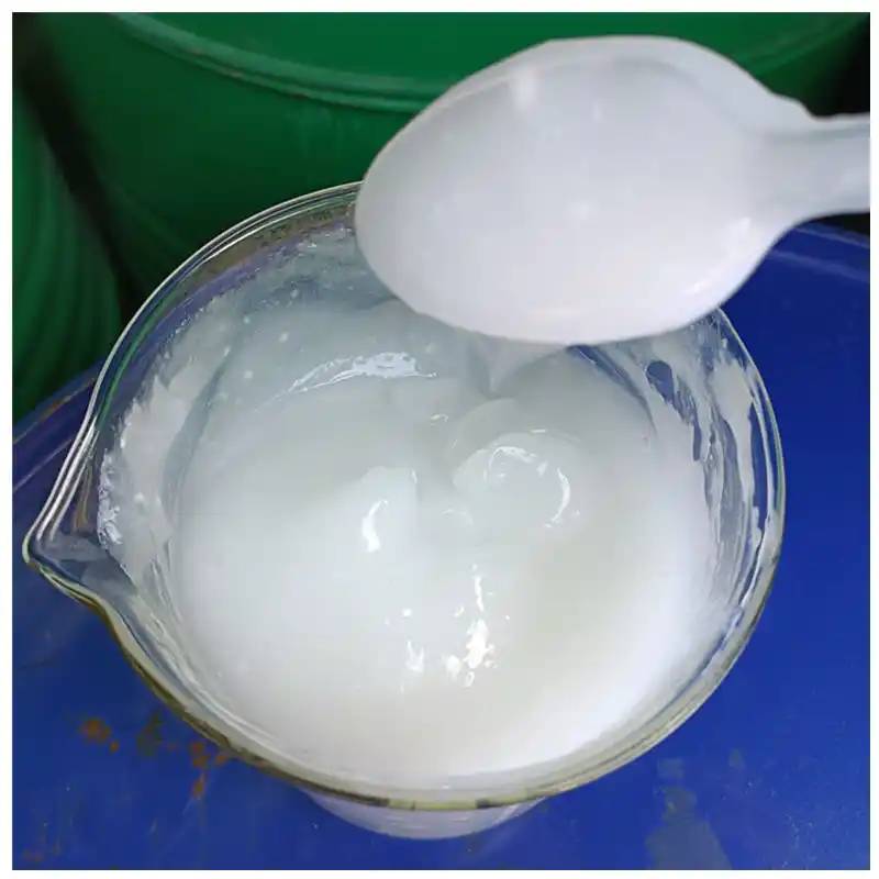
Hyaluronic acid is a water-retaining polymer that is naturally found in human skin. It is formed by the repeated alternation of N-acetylglucosamine and glucuronic acid disaccharide units. The highly extended and randomly coiled conformation of the hyaluronic acid molecule in solution allows it to occupy a large area, and the molecular chains are intertwined to form a continuous network structure. Water molecules interact directly with it through polar bonds and hydrogen bonds, making hyaluronic acid act like a molecular sponge, absorbing and holding thousands of times its own weight in moisture. It is internationally recognized as the best moisturizer[2] and is known as the “natural moisturizing factor” (NMF). In connective tissue, the water retention value of hyaluronic acid is about 80 ml/g. It has a stronger water retention capacity than other natural or synthetic polymers. Some scholars [3] compared the water retention properties of hyaluronic acid with those of sorbitol and polyethylene glycol 4000. The results (hyaluronic acid molecular weight 3000kD), sorbitol, and polyethylene glycol 4000 24h water retention were 87.1, 32.4, and 10.5, respectively. The water retention of hyaluronic acid is one of its most important physiological functions.

1.2 Hyaluronic acid participates in the formation of proteoglycan polymers
Under physiological conditions, hyaluronic acid is covalently linked to core proteins in concert with other glycosaminoglycans (such as chondroitin sulfate and keratan sulfate) to form proteoglycan polymers. These giant polymers occupy a large water pressure domain, and can reduce in volume under external pressure. After the pressure is released, they expand to their original volume, thereby maintaining the shape and volume of the tissue and ensuring its reversible compressive resistance. Proteoglycans (PGs) in different tissues have their own special functions. For example, the proteoglycans distributed in connective tissue bind water through amino sugars. This hyaluronic acid-protein-water gel binds cells together, enables normal cell metabolism and tissue water retention, and protects cells from pathogenic bacteria to prevent infection. and gives the skin a certain elasticity. In cartilage, it determines the volume of cartilage and the final shape of the skeleton. In the aorta, it is essential for the optical physiology of the eye together with the vitreous body. In the ovary, PG (without hyaluronic acid) is synthesized regularly with the periodic growth of follicles. The heparan sulfate proteoglycan on the plasma membrane of human liver cells plays an important role in the interaction between cells and the interaction between cells and the matrix.
1.3 Hyaluronic acid: Effects on cell biology
Toole et al. demonstrated that hyaluronic acid is highly abundant during embryogenesis and morphogenesis, and that during differentiation, hyaluronidase removes this hyaluronic acid, producing a differentiated matrix, such as proteoglycans and collagens [4]. The role of hyaluronic acid during differentiation is to cause isotonic tissue edema, which leads to the opening of channels required for cell migration; to assist in the separation and transposition of mesenchymal cells released from the epithelium; and to change the shape and structure of organs.
1.4 The role of hyaluronic acid on immune cells
Domestic and foreign studies have confirmed that hyaluronic acid has a certain effect on macrophages, granulocytes, lymphocytes, natural killer (NK) cells, etc. Hyaluronic acid at low concentrations has a mild effect on the function of phagocytes and NK cells, but inhibits the transformation of lymphocytes and the formation of E rosettes. Hyaluronic acid at higher concentrations has a significant inhibitory effect on the function of lymphocytes, NK cells and phagocytes, and there is a dose-effect relationship.
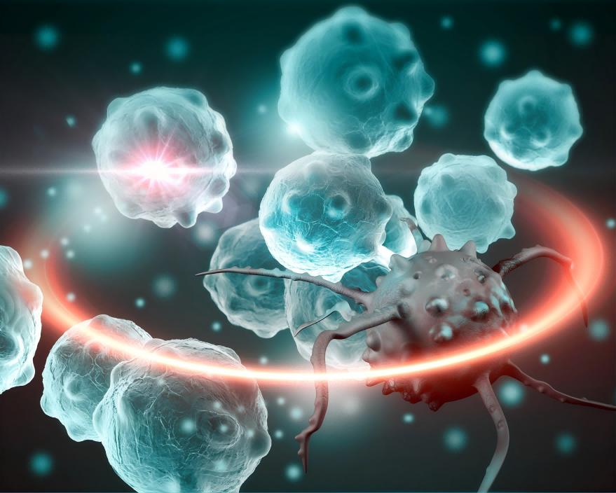
The extracellular coat (Coat) formed by hyaluronic acid acts as a barrier against cytotoxic lysis. For example, the glycosaminoglycan coat of glioma cells can enhance their resistance to immune killing cells; the hyaluronic acid coat adhering to the cells can block lymphocyte-mediated cell lysis. In addition, this hyaluronic acid coat can prevent viral invasion (such as the invasion of the Newcastle disease virus into human synoviocytes), and interfere with the function of intercellular lectins and lectin receptors.
1.5 Effect of hyaluronic acid on wound healing
Hyaluronic acid can accelerate wound healing. Local hyaluronic acid concentration increases significantly in the early stages of a wound, and it has a regulatory effect on a variety of inflammatory cells. High-concentration, high-molecular-weight hyaluronic acid has a higher pro-healing effect, regulates the synthesis of collagen, regulates fibrous activity and has anti-inflammatory effects. Low-molecular-weight hyaluronic acid, on the other hand, stimulates angiogenesis. Hyaluronic acid in wounds is broken down by enzymes into low molecular weight hyaluronic acid, which in turn promotes wound healing.
2 Applications of hyaluronic acid
2.1 Hyaluronic acid in cosmetics
2.1.1 Hyaluronic acid and phospholipids form an emulsifier [5]
In the absence of other emulsifiers, the addition of hyaluronic acid and phospholipids to an oil-water mixture can form a stable emulsion. This natural emulsifier composed of hyaluronic acid and phospholipids is characterized by both emulsifying and moisturizing properties. It is a safe and effective emulsifier that cannot be matched by other synthetic surfactants and can be used in the preparation of skin creams, lotions, facial cleansers, etc.
2.1.2 Hyaluronic acid and polyoxyethylene form a thickener [6]
Polyoxyethylene (molecular weight 100-5000kD) is a commonly used thickener in cosmetics, and hyaluronic acid (molecular weight 3000kD) solution also has a high viscosity. The viscosity of the two combined is much greater than the sum of their individual viscosities. Therefore, hyaluronic acid-polyoxyethylene solution is an excellent thickener with good moisturizing properties, and can be used in cosmetics such as creams and lotions.
2.1.3 Hyaluronic acid as a fragrance fixative [7]
According to the characteristics of hyaluronic acid, which has the function of molecular encapsulation of various substances, it is used in fragrance products. Hyaluronic acid, as a fixative combined with fragrance, can slow down the volatilization rate of the fragrance and make the fragrance last longer. It is suitable for perfumes, skin care creams, emollients, deodorants, etc. Hyaluronic acid has two advantages in addition to fixing the fragrance: first, it reduces the adverse irritant effect of the fragrance on sensitive skin and reduces allergic reactions; second, it prevents adverse chemical reactions between certain fragrances and skin secretions and prevents the formation of odors.

2.2 Value of hyaluronic acid in clinical diagnostics
Hyaluronic acid levels are significantly elevated in many diseases. Clinically, measuring the serum hyaluronic acid level can reflect changes in various diseases. Studies have shown that changes in serum hyaluronic acid levels are closely related to the course of liver disease and the degree of liver cell damage. In recent years, domestic and foreign scholars have confirmed that the serum hyaluronic acid of patients with liver cirrhosis is significantly elevated, and the degree of elevation of hyaluronic acid is positively correlated with the degree of liver cirrhosis. Therefore, serum hyaluronic acid can be used as a reliable indicator for the diagnosis of liver cirrhosis. Presig believes that this indicator is significantly superior to previous indicators for diagnosing liver cirrhosis (such as the albumin/globulin ratio, pro-collagen III peptide, etc.), and can better reflect the degree of liver cirrhosis than liver puncture and biopsy.
2.3 Clinical value of pure hyaluronic acid products
In the 1980s, Balazs and others developed a cross-linked hyaluronic acid derivative, hylan, and explored its physiological properties and clinical applications. It was found that hylan has the same biocompatibility as natural hyaluronic acid, inspiring people to clinically prevent adhesion after trauma or surgery and repair soft tissues [8].
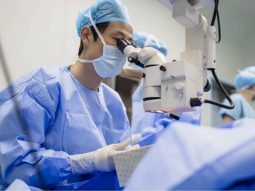
2.3.1 Hyaluronic acid in ophthalmic surgery [9,10]
Hyaluronic acid gel and its high elasticity can be used in a variety of ophthalmic microsurgery. It has been recognized that hyaluronic acid gel is essential in ophthalmic microsurgery. Hyaluronic acid gel is now widely used as an essential filler in various ophthalmic surgeries such as extracapsular cataract extraction, intraocular lens implantation, retinal detachment surgery and corneal transplantation. In addition, hyaluronic acid can also be used for eye canal reopening and dry eye syndrome. Extensive clinical applications over the past 20 years have shown that the role of hyaluronic acid viscoelastic substances in ophthalmic microsurgery is not simply to provide operating space, but also to provide viscoelasticity without a lining, tissue separation, soft tissue restoration, viscous occlusion, viscous bleeding, viscoelastic buffering and viscoelastic fixation, which can greatly reduce surgical trauma, reduce the degree of postoperative inflammatory response, reduce surgical complications, and achieve the goal of improving vision.
2.3.2 Hyaluronic acid in the treatment of bone and joint diseases
The lubricating and cushioning effect of hyaluronic acid has led to its use in the treatment of osteoarthritis, periarthritis of the shoulder and rheumatoid arthritis. Hyaluronic acid gel is used for intra-articular injections to treat osteoarthritis. Not only does it overcome the side effects of previous hormone treatments, but its short- and long-term effects are also superior to those of hormones, making it the material of choice for treating osteoarthritis. At the 18th International Anti-Rheumatic Federation meeting in July 1993, hyaluronic acid was considered to have a disease-repairing effect on osteoarthritis and is a disease-repairing osteoarthritis treatment [11]. In fact, since hyaluronic acid injections for osteoarthritis were marketed in Italy in 1987, intra-articular injections have been marketed in many countries, and clinical trials are also being conducted in the UK and the US. Japan Bio-Chem's hyaluronic acid injections for osteoarthritis improved symptoms by 64.1%, and sales of the product reached 27.1 billion yen in 1993 [12].
2.3.3 The application of hyaluronic acid in tympanic membrane repair [13]
Tympanic membrane perforation is a common clinical condition caused by acute and chronic otitis media, trauma, etc. Surgical repair of the tympanic membrane often causes changes in the structure of the original tympanic membrane after surgery, affecting the degree of hearing recovery. This is mainly related to the formation of scar tissue and the use of graft materials. In recent years, domestic and foreign research on the application of medical sodium hyaluronate in tympanic membrane repair has achieved gratifying results. Summary: A large number of clinical results show that hyaluronic acid treatment for tympanic membrane perforations is feasible, without any side effects, and is a good means of treating small dry tympanic membrane perforations.
2.3.4 Hyaluronic acid: Clinical application in obstetrics and gynecology
(1) A: Application after laparoscopy, hysteroscopy and other intrauterine operations
It is known in medical practice that hyaluronic acid is present in the peritoneal fluid, uterine cavity fluid and fallopian tube fluid. The use of hysteroscopy and laparoscopy can cause the loss or dilution of components of the abdominal or uterine cavity fluid, and temporary real space gaps in hyaluronic acid may also occur. Local trauma, bleeding and exudation during surgery can disrupt the internal environment of the uterus, affecting the function of the female reproductive tract and mucosa to varying degrees. The interaction of these factors can lead to impaired organ function or infertility and other consequences. Hylan B gel is a highly viscoelastic. A non-permeable derivative, its polysaccharide chains are cross-linked by divinyl sulfone, making it a good soft tissue filling material. Therefore, in laparoscopic and hysteroscopic use, Hylan B gel can be used as an operating medium for the lens to separate tissues and expose the field of view, which not only accelerates the repair of the damaged surface and the amount of synthesis, but also does not affect the surgical field of view, facilitating operation and reducing the possibility of postoperative complications such as blockages and adhesions. [14]
(2) The application of hyaluronic acid in obstetrics and gynecology to prevent postoperative adhesion
In 1971, Balazs et al. reported the role of hyaluronic acid in preventing adhesion formation between tendons and sheaths, conjunctiva and iris. They found that high viscoelastic, high molecular weight hyaluronic acid solutions and hyaluronic acid membranes can reduce the incidence of adhesion formation, and have no adverse effect on wound healing. A large number of animal experiments and clinical applications have shown that hyaluronic acid is a safe and effective substance for preventing and reducing adhesion caused by surgery.
(3) Hyaluronic acid in the vagina
Hyaluronic acid is secreted and distributed in the fallopian tubes and guiding tissues of mammals, as well as between the epithelial cells of the mucous membrane. Experiments have found that low-density areas of vigorous cell proliferation are always accompanied by increased synthesis of hyaluronic acid. Hyaluronic acid and its derivatives act as a medium for controlled drug release. Therefore, in clinical use of vaginal medication, the moisturizing and lubricating properties, viscoelasticity, and characteristics of hyaluronic acid as a drug carrier can be utilized. Hyaluronic acid can both increase the self-cleaning and lubricating effects that have been disrupted in the vagina, allowing the local epithelial cells to recover, while also slowly releasing the drug it is carrying into the vagina, where it can exert a pharmacological effect for a long time, restore the vagina to its normal state, and heal the original disease.
(4) Extended use of hyaluronic acid preparations
Wound healing is a process involving multiple cells and their products that work together to rebuild and regenerate the extracellular matrix. Current research has confirmed that hyaluronic acid plays an important role in the process of wound healing. With the development of fetal surgery, a glycoprotein called hyaluronic acid stimulatory factor has received increasing attention. It can significantly increase the hyaluronic acid content in the wound matrix, thereby regulating collagen synthesis. This is considered to be an important factor in the fetal wound healing process without scarring.
2.4 Discussion of the safety of hyaluronic acid
The hyaluronic acid in the preparation is the same as the endogenous hyaluronic acid, which is non-toxic, non-antigenic, and not likely to cause a foreign body reaction. Clinical applications have shown that hyaluronic acid has good safety, and most of them indicate that hyaluronic acid is well tolerated, with an adverse reaction rate of 0% to 10%. Japan has conducted comprehensive preclinical and clinical safety experiments on hyaluronic acid preparations, which have proven their safety and efficacy in clinical use. Common adverse reactions are mostly mild to moderate pain or swelling at the administration site or injection site, as well as symptoms such as headache or fever in individual patients. Adverse reactions often occur within 1 to 2 days of administration, and patients can generally tolerate them without treatment, and they will disappear on their own after 2 days. Obstetrics and gynecology, especially when used in the vagina, has very low absorption, and hyaluronic acid has even fewer adverse reactions. Currently, hyaluronic acid macromolecular cross-linked membrane agents and compound preparations with other anti-adhesion drugs are being studied for their potential in anti-adhesion due to their good safety and controlled-release effects.
3 Production of hyaluronic acid
3.1 Extraction from animal organs
Hyaluronic acid is mainly extracted from umbilical cords, cockscombs, vitreous bodies of cattle and sheep eyes, and whale cartilage. These animal tissue sources are difficult and expensive to obtain, and the hyaluronic acid content in the tissue is very low, resulting in a low yield of product. During the extraction process, large amounts of organic solvents and enzymes must be used, and the process is complex and involves many operating units, which greatly increases the cost of hyaluronic acid and makes it difficult to produce it on a large scale. Foreign companies that use this method include Kabi, Fidia, Gen-zyme, Sweden's P-hyaluronic acidrmacia and Japan's Biochemical Industry Company. The level of research in China is relatively low, and hyaluronic acid production is still in the organ extraction stage, with the main raw materials being umbilical cords and cockscombs. The Shanghai University Journal reported a method for extracting hyaluronic acid from pig skin, and the molecular weight of the resulting hyaluronic acid is about 106. Currently, companies that use this method to produce hyaluronic acid include Shandong Freda Pharmaceutical Company and Shaanxi Pharmaceutical Industry Research Institute.
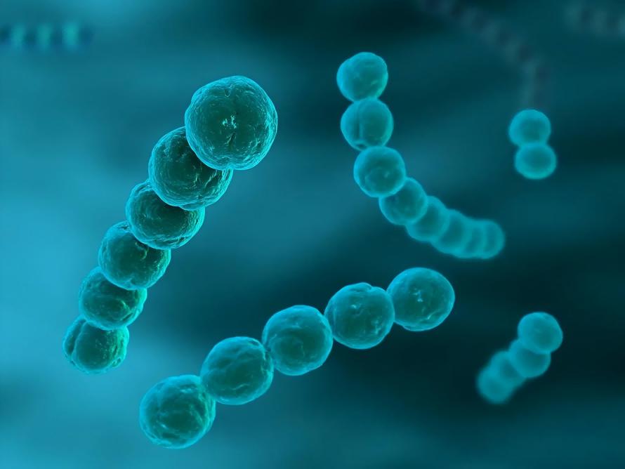
3.2 Microbial fermentation method
As early as 1937, Kendall et al. [15] discovered that streptococci could produce hyaluronic acid, and many people have conducted research on this. Shiseido of Japan first reported the use of streptococci to produce hyaluronic acid in 1985. China is currently developing this method, and Yan Jialin of Zhengzhou Animal Husbandry Engineering College and others have used hyaluronic acid-deficient streptococci to ferment and produce hyaluronic acid that meets cosmetic requirements. Strains with high fermentation yields are generally obtained by mutagenesis. A Japanese patent reports a method of mutagenesis using NTG (N-methyl-N-nitro-nitrosoguanidine), and a two-step mutagenesis treatment is used to obtain a variant that does not produce hyaluronic acid.
The fermentation rate is as high as 6.7 g/l, and the molecular weight is ≥10×105. The fermentation yield of the original strain without mutagenesis is only 2 g/l, and the molecular weight is 3×10⁵ to 6×10⁵. The mutagenesis rates of the two-step treatment are 4×10⁻⁶ and 5×10⁻⁵, respectively. In other words, it takes hundreds of thousands or even millions of bacteria to obtain a desired variant, and the amount of work involved is imaginable. A new fermentation process for producing hyaluronic acid has also been developed in China, using hyaluronic acid-producing bacteria obtained by gamma irradiation combined with magnetic field mutagenesis. The results passed the national-level achievement appraisal organized by the Ministry of Chemical Industry on July 29, 1998, and the conclusion was that the technical level has reached the international advanced level. The fermentation level of hyaluronic acid is 4-6.5 g/l. According to literature, the current international fermentation level of hyaluronic acid is 6.5 g/l at the highest.
3.3 Animal fetal cell separation method
The American biotechnology company Genzyme made this a key topic in 1994, attempting to replace the current method of extracting from animal organs. That is, in an animal cell culture, a certain type of cell is propagated in large quantities to extract a certain substance, which is an important field of biotechnology [16]. However, there are currently no documented or reported cases of this method.
References:
[1] Meyerk, Palmer Jw. The polysacc hyaluronic acidride of the vitreous houmor [J]. J Biolchem, 1934, (107): 629-633.
[2] Wu Dongru. Biochemistry of carbohydrates [M]. Beijing: Higher Education Press, 1987: 627.
[3] Nagai Masayoshi. Cosmetic ingredients. JP Show 62-158203, 1987.
[4] Zhang Shimin, Zhang Lurong. Chemistry of life [J]. 1991, (11) 2: 8-9.
[5] Ujiri Yoshitaka. Emulsifying composition. JP Show 62-45336, 1987.
[6] Xu Hong, Lu Zhihua. Chinese Journal of Biochemical Drugs [J]. 1998, (19) 5: 222-223.
[7] Bracke Jw. Hyaluronic acid/hyaluronate tased fragrance products, WO 85/04803, 1985.
[8] Hou Junji, Zhang Tianmin, Niu Guizhen. Chinese Journal of Biochemical Drugs [J]. 2003, (24) 5: 262-264.
[9] Zhou Libin, Zhang Daqin. Chinese Journal of Biochemical Drugs [J]. 1998, (5): 289.
[10] Zhang Yanbing, Tian Yeqing. Chinese Journal of Biochemical Drugs [J]. 1998, (5): 288.
[11] Li Weiping, Liu Shangli, Lin Daoxian. Chinese Journal of Biochemical Drugs [J]. 1998, (5): 252-254.
[12] Luo Ruiming. Journal of Ningxia Agricultural College [J]. 2001, (22) 1: 62-64.
[13] Piacquadio DJ, Larsen NE, Denlinger JL, et al. Hylan B gel (hylaform) as a soft tissue augmentation material [A]. Klein AW. Tissue-augmentation in clinical practice: procedures and techniques [C]. New York: Marcel Dekker, 1998. 269-291.
[14] Yang Xiaohong, Ling Peixue, Wang Fengshan. Chinese Journal of Biochemical Drugs [J]. 1998, 19 (4): 205-208.
[15] Kendal PE. A serologically inactive polysacc hyaluronic acidride elaboraled by mucoid strains of group A strcptococcus [J]. J Biol Chem, 1937, (118): 61-69.
[16] Xu Jien. Jiangsu Food and Fermentation [J]. 1996, (1): 27-29.
Recommend Information
-
What Is the Benefit of Low Molecular Weight Hyaluronic Acid Powder?

-
What Are the Preparation Methods of Low Molecular Weight Hyaluronic Acid Powder?

-
Research on the Structure, Performance, Modification and Application of Hyaluronic Acid

-
How to Produce Hyaluronic Acid Powder by the Fermentation Method?


