English
 French
 Spanish
 Russian
 Korean
 Japanese
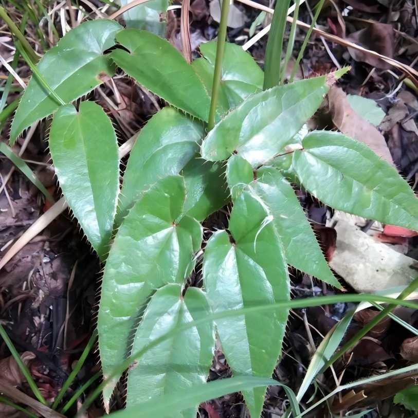
Natural Feed Additive Epimedium Extract Promote Healthy Breeding
Epimedium (Epimedium L.) is a genus of herbaceous plants rich in resources within the Berberidaceae family. China is home to 57 endemic species of this genus, which are widely distributed in Shaanxi, Liaoning, Guangxi, Hubei, Sichuan, and other regions, with stable sources and diverse varieties.The whole plant contains various components, such as Epimedium total flavonoids, Icariin, Epimedium polysaccharides, terpenoids, alkaloids, lignans, chlorogenic acid, and phytosterols, exhibiting excellent natural properties and safety.
In modern livestock farming, natural plant-based raw materials are increasingly valued for their green and sustainable characteristics. As a professional supplier of plant extracts, Green Spring Technology leverages its comprehensive supply chain and stringent quality control system to provide customers with high-purity, high-stability Epimedium extract raw material solutions. We are committed to helping customers:
· Enhance product competitiveness: Provide compliant, traceable natural raw materials to meet market demand for green, safe feed additives;
· Optimise animal growth performance: Support customers in developing scientific formulations to enhance animal health and production performance;
· Meet industry regulatory requirements: All products comply with national standards for feed raw materials, ensuring the legal and safe market release of customer products.
Green Spring Technology provides stable and high-quality Epimedium extract solutions to livestock, feed, and farming enterprises with professional expertise and reliable supply, collaborating with customers to drive the industry towards a natural, efficient, and sustainable future.
1 Main Components of Epimedium Extract
1.1 Core Component of Epimedium Extract: Flavonoids
In the field of natural plant-based raw materials, Epimedium has garnered significant attention due to its rich active components. Among these, flavonoids, as one of its core components, have become a key focus for product development and quality control at Green Spring Technology.
Flavonoids are natural compounds composed of two phenolic hydroxybenzene rings (A ring and B ring) connected by a three-carbon bond. They are abundant and diverse in Epimedium. Research has identified over 21 flavonoid compounds in Epimedium, including epimedium glycoside, epimedium A, epimedium B, and epimedium C. The discovery of new compounds such as 3-hydroxybaicalin-II has opened up new possibilities for product development.
As a professional plant-based raw material supplier, Green Spring Technology places great emphasis on the source variability and component stability of raw materials. We have found that the flavonoid content in Epimedium varies significantly depending on the variety, part, and origin of the plant: Epimedium leaves from Korea contain higher levels of epimedium glycoside and total flavonoids, while the rhizomes are richer in epimedium C ; Habitat influence is also pronounced—in secondary broadleaf forests, Icariin content can reach 1.37%, and total flavonoids up to 18.40%, significantly higher than in pine forests.
Green Spring Technology relies on standardised cultivation bases and a strict raw material procurement system to ensure that we provide customers with high-quality Epimedium extracts with stable and consistent batches of flavonoid components such as quercetin, kaempferol, luteolin, and epimedium glycoside. In the future, we will continue to deepen component research and process innovation, collaborating with industry partners to promote the application and development of natural plant raw materials in the health product sector.

1.2 Core Component of Epimedium Extract: Polysaccharides
Polysaccharides are an important functional component in Epimedium, with their content and composition significantly dependent on the variety origin, growing environment, and plant parts used. Through systematic research and technological integration, Green Spring Technology has established a comprehensive supply chain for Epimedium polysaccharide raw materials, capable of providing various refined components, including neutral polysaccharides and acidic polysaccharides.
These components are composed of various monosaccharides such as galactose, glucose, arabinose, xylose, rhamnose, mannose, and glucuronic acid, exhibiting clear structural composition and stable content characteristics.
During product development, Green Spring Technology places particular emphasis on the consistency and traceability of raw materials. Research has shown that the polysaccharide content in the roots of Korean Epimedium is relatively high, with a total content range of 22.47% to 31.11%, and that the variety and geographical origin of the plant significantly influence polysaccharide yield. Based on this finding, the company has optimised raw material selection and process control to achieve precise utilisation of Epimedium raw materials from different varieties and plant parts, ensuring high batch-to-batch quality consistency of the final extract products.
In the future, Green Spring Technology will continue to advance research on the application of Epimedium polysaccharide components in new raw material fields, collaborating with industry partners to develop natural, trustworthy, and efficient plant-based raw material solutions.
1.3 Core Component Three of Epimedium Extract: Lignans and Alkaloids
Lignans are a class of natural organic compounds formed by C6-C3 units connected via side chain β-carbon atoms. Epimedium contains various lignan components, primarily including (+)-cycloartemisin, cyperin A, (+)-cyperin, (+)-isocyperin, and cyperin C, among others [14].
Ti et al. [15] isolated new 8-O-4'-type lignan components from Epimedium pseudowushani. Alkaloids are another class of components in Epimedium, with three alkaloid components currently identified and isolated from Epimedium, the main component being magnolol.
The content of magnolol varies among different Epimedium species: the highest content was found in Korean Epimedium, followed by rough-haired Epimedium and Tianping Mountain Epimedium, with content generally exceeding 0.07% [16]. The distribution of magnolol also varies among different plant parts, with higher content in roots and stems than in leaves [17].
2 Application of Epimedium and its extracts in chicken production
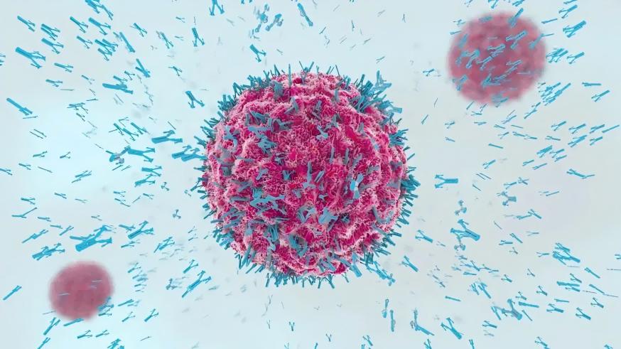
2.1 Effects on chicken immune performance
Epimedium extracts, as a plant-based feed ingredient, have increasingly attracted attention for their potential applications in chicken production.
Their main components, such as Icariin and epimedium polysaccharides, have been observed to be associated with immune parameters and intestinal structure in chickens in relevant studies. For example, studies have shown that adding a certain amount of Icariin to the diet may be associated with the secretion trends of interleukin-2 (IL-2) and interferon-γ (IFN-γ), while levels of Newcastle disease virus (NDV) antibodies and globulin also exhibit changes. Another study indicated that EPS-1, a polysaccharide from Epimedium, demonstrated potential effects on cell activity in vitro and may help maintain healthy levels of blood coagulation inhibitory antibodies and lymphocyte status in chickens under rearing conditions.
Regarding intestinal health, experiments have shown that adding Epimedium extract to drinking water may have a positive impact on the tissue morphology of the caecal tonsils. The phased use of EPS-1 has also been found to be associated with adjustments in the villus morphology of broiler chickens.
In summary, Epimedium extract, as a feed ingredient, may help support the healthy growth of chicken flocks and provide a new nutritional support option for poultry production.
2.2 Epimedium extract enhances egg production performance and egg quality in laying hens
As a natural plant-based feed ingredient, the application value of Epimedium extract in layer chicken nutrition has gradually emerged in recent years. Its various active components help optimise feed composition, promote nutrient absorption, and have positive effects on layer chicken production performance and egg quality.
Studies have shown that adding 0.20% Icariin (ICA) to layer chicken diets helps increase average egg weight and improve protein content and Haugh units during the later stages of feeding. Additionally, experiments have found that incorporating plant additives containing Epimedium into the diet of Guifei chickens significantly reduces egg yolk cholesterol levels at different time points while improving overall egg production performance. Related studies also indicate that daily supplementation with appropriate amounts of compound plant materials containing Epimedium can increase egg production rate and average daily egg weight, while significantly optimising the feed-to-egg ratio.Furthermore, adding 0.20% ICA to the diet was observed to help increase the average daily feed intake of laying hens.
In summary, Epimedium extract can serve as an effective feed ingredient, supporting the healthy rearing of laying hens and improving egg quality by enhancing average egg weight, protein quality, cholesterol content, and feed conversion efficiency.
2.3 Application and Potential of Epimedium Extract in Chicken Antioxidant Function
In modern farming, chicken flocks often face challenges related to oxidative stress. Epimedium extract, as a natural plant-based feed ingredient, contains active components such as epimedium glycoside and epimedium glycoside II, which exhibit good antioxidant potential and help maintain overall health.
Studies have shown that adding 0.20% epimedium glycoside (ICA) to the diet of laying hens significantly reduces malondialdehyde (MDA) levels in oviduct tissues, suggesting its potential to mitigate lipid peroxidation. The use of epimedium polysaccharides in the chick stage has also been observed to increase the activity of various antioxidant enzymes (such as SOD, GR, GSH-Px, and CAT) in serum while reducing MDA levels, with the 25 mg/mL treatment group showing the most prominent effects.
Additionally, some compound plant materials containing epimedium have been found to potentially influence the expression of genes related to antioxidants.
In summary, Epimedium extract can help maintain the redox balance in chickens through multiple pathways, providing further support for poultry health and farming.
3 Green Spring Technology's high-quality Epimedium extract offers a new option for poultry health and farming
Epimedium, a natural plant rich in resources and diverse active components, is gaining increasing attention in chicken production.
Current research primarily focuses on components such as Icariin and epimedium polysaccharides. However, Green Spring Technology, leveraging its continuous R&D and technical expertise, is actively advancing the development of more active components from epimedium and their application in feed, offering customers more scientifically formulated solutions.
In practical applications, Epimedium extract is often used in combination with other plant components. Through systematic research and formulation experiments, Green Spring Technology has developed several compound products that better meet the needs of different farming scenarios. Additionally, for the appropriate addition levels of Epimedium extract at different growth stages of chickens, Green Spring Technology provides detailed data and comprehensive application guidance to help customers achieve scientific feeding, cost reduction, and efficiency improvement.
Green Spring Technology supplies Epimedium extract in various specifications (including Icariin at 10%-98% and other customised gradients), with stable purity, strong batch-to-batch consistency, and complete compliance certifications and testing reports. This helps customers precisely control additive dosages, optimise formulation costs, and effectively enhance the stress resistance and overall production performance of chicken flocks.

Visit Green Spring Technology's official website or contact our customer service team at helen@greenspringbio.com or WhatsApp: +86 13649243917 to request free samples and technical documentation. Let's collaborate to harness the power of natural plants and create greater value for healthy livestock farming!
References
[1] Ye Lika, Chen Jimin, Liu Sihai, et al. Pharmacokinetics of icariin in rats [J]. Chinese Journal of Pharmacy, 1999, 34 (1): 33.
[2] Zou Jieming, Meng Jie, Yan Zhenghua, et al. Pharmacokinetic study of the active ingredient icariin in the traditional Chinese medicine compound Epimedium [J]. Traditional Chinese Medicine, 2002, 33 (1): 55.
[3] Liu Tiehan, Wang Yi, Wang Benxiang, et al. Study on the intestinal bacterial metabolism of icariin I. Metabolic transformation of icariin by intestinal bacteria [J]. Traditional Chinese Medicine, 2000, 31 (11): 834.
[4] Ge Linfu, Dong Zhengjun, Jiang Guosheng, et al. The effect of icariin on the proliferation and differentiation of drug-resistant and non-drug-resistant HL-60 cells [J]. Chinese Journal of Practical Medicine, 2001, 3(3): 25.
[5] Bi Kehong, Zhang Yukun, Ge Linfu, et al. Effects of icariin on the immune and hematopoietic functions of irradiated mice [J]. Chinese Journal of Radiation Hygiene, 2001, 10(2): 104.
[6] Li Shutong, Li Tiejun, Zheng Yueqin, et al. Effects of icariin on the function of peritoneal macrophages in mice [J]. Journal of the Second Military Medical University, 1995, 16(6): 541.
-
Prev
What Are the Benefits of Black Garlic Extract?
-
Next
Green Spring Technology's Multi-Specification Epimedium Extract Icariin Helps Customers Develop Products Efficiently