English
 French
 Spanish
 Russian
 Korean
 Japanese
What Is Natural Coloring?
Natural coloring has a wide range of sources and a wide variety of types in nature. Natural coloring from different sources may also have very different molecular structures. The first condition for applying natural coloring is to explore, classify and summarize its structure. In practice, the primary challenge in the practical application of natural coloring is how to achieve efficient and low-cost extraction while maintaining the original color and function of natural coloring. With the development of technology, the extraction of natural pigments has gradually evolved from the most primitive methods such as the pulverization method and the maceration and pressing method to modern, low-cost, high-efficiency, and intelligent techniques such as the solvent extraction method, the enzymatic hydrolysis method, the supercritical fluid extraction method (SCF), and the high-pressure pulsed electric field method (PEF). However, Natural Coloring itself is unstable, and how to improve its stability in different environments while maintaining its color and performance is also a major problem that needs to be solved in the practical application of Natural Coloring.
From the perspective of application scenarios, Natural Coloring is mainly used to replace synthetic coloring in industries such as food and printing. In addition, some natural pigments are often specially treated and used in industries such as healthcare, solar cells and intelligent detection because of their properties of promoting human health, anti-cancer, suitable for preparing photosensitizers, and changing color in different environments.
1 Sources of Natural Coloring
1. 1 Plant Based Natural Coloring
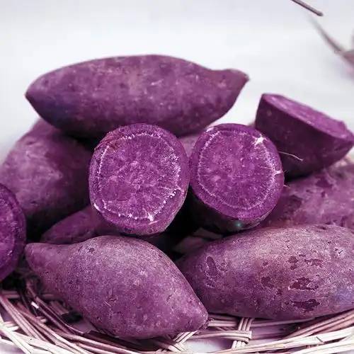
Plant-based natural coloring refers to natural pigments produced by the metabolism of plants themselves. These pigments not only give plants different colors, but also play an important role in the life activities of plant bodies. Different types of pigments are distributed in different parts of the plant. For example, as early as 1815, German scientists Vogel and Pelletier had already isolated the curcumin molecule from the rhizomes of turmeric [1]. Among plant-derived natural colorings, anthocyanins are important active substances for coloring flowers and fruits. They include more than 700 polyphenolic pigments in the flavonoid family [2]. Zhang et al. [3] extracted anthocyanins from purple sweet potatoes and red leaf cabbage in an ethanol solution, mixed them with corn starch and PVA to prepare a freshness indicator film, and used the film to detect the freshness of shrimp by color change.
1.2 Animal-derived Natural Coloring
Animal-derived Natural Coloring is produced by animals to ensure their normal physiological needs and protective functions. It is mainly in the form of three types of pigments: porphyrin pigments, polyene pigments, and indole pigments. Common examples are melanin containing phenols or indoles, heme from porphyrin pigments, and astaxanthin from polyene pigments.
1.3 Microbial sources Natural Coloring
Common microorganisms such as fungi, bacteria and microalgae also produce natural pigments. The Natural Coloring produced by different microorganisms differs significantly in terms of chemical composition, stability, solubility and function. The main pigments from microbial sources that have been reported are riboflavin, β-carotene, canthaxanthin, spirulina red, phycobilin, melanin, violacein, astaxanthin and lycopene [4]. For more information, see Table 1. Most of them are used as food colors.
2 Classification of Natural Coloring
Natural Coloring is mainly divided into five categories, as shown in Table 2.
3 Extraction of Natural Coloring
The most common extraction methods for Natural Coloring include the crushing method and the maceration and pressing method [8], but these methods have disadvantages such as low extraction rates and low product purity. To achieve the widespread and effective application of Natural Coloring, the improvement of its extraction methods is essential.
3. 1 Solvent extraction method
Solvent extraction is one of the simplest and most widely used methods for pigment extraction. Organic solvents such as acetone, methanol, isopropanol and petroleum ether can all be used as extraction solvents. Each solvent has a different polarity, and the most suitable polar solvent can be selected according to the polarity of the pigment. Zhao et al. [9] found that acetone was the most suitable solvent for extracting natural astaxanthin [10], with a recovery rate of about (44± 1)%. This may be because the structure of acetone contains many carbonyl groups that are highly similar to natural astaxanthin.
Microwave-assisted extraction technology is a new extraction technology that can achieve instantaneous penetration heating based on traditional solvent extraction. This method has strong penetration into various container materials, and has a wide range of selectivity for the extracted organic solvents and the extracted materials, as well as a high temperature rise efficiency. Under the action of microwave radiation, plant cell tissues will produce rapid and irregular intermolecular movements, This movement can cause friction within the substance and generate heat energy, which causes the cell walls and tissues to break and the solutes to flow out, accelerating the extraction rate and effectively increasing the yield of the product [11-12]. Suntaro and Tsubaki et al. [13] demonstrated the effectiveness of microwave-assisted hydrothermal extraction for the extraction of sulphated polysaccharides from daylily, daylily tuberose and daylily broadleaf.
Ultrasonic extraction (UAE) is a kind of assisted extraction method with low temperature extraction (40-60°C), non-slow extraction (20-40 min), and high extraction efficiency (increased by more than 50%). The method generates ultrasound that can shuttle and vibrate in the liquid solvent, producing ultrasound “explosion” and promoting the production of natural coloring [14]. Zhu et al. [15] extracted natural anthocyanins from purple sweet potatoes using UAE and studied the process conditions with the highest extraction content: ultrasound time of 40 min, assisted thermal extraction at 80 °C, pH of 2.5, and ethanol concentration of 58%. The anthocyanin and non-anthocyanin content of purple sweet potatoes extracted using UAE was higher than that of the conventional method, indicating that UAE effectively improves the extraction of anthocyanins from purple sweet potatoes.
Accelerated solvent extraction (ASE) is widely regarded as an extraction technique that is extremely suitable for polar compounds [16-17]. The method was first reported in 1996. Its core technology is to use ultra-high temperatures (up to 200 °C) and ultra-high pressures (1000–3000 psi or 10.3–20.6 MPa) to achieve rapid (about 5–20 min) extraction of solid and semi-solid samples [18].
ASE is an excellent choice for extracting natural products with poor thermal stability. The short extraction time at high temperature and pressure is very beneficial for the protection of Natural Coloring components that are less thermally stable. Cai et al. [19] studied the extraction efficiency of anthocyanins by conventional extraction, UAE and ASE, and found that the extraction efficiencies of these three methods for anthocyanins were in the following order: ASE>UAE>conventional extraction. In terms of anthocyanins, compared with conventional extraction and UAE, ASE extracted more diacyanidin, and less nonacyl and monoacyl anthocyanins, which also fully demonstrates that the anthocyanins extracted by ASE are more stable than those extracted by conventional extraction and UAE. Maja et al. [20] confirmed through ASE that wild nettles are rich in bioactive molecules such as low molecular weight polyphenols and pigments, including 41 phenolic compounds, 13 carotenoids and 9 chlorophylls. Truong et al. used ASE as an extraction method for different genotypes of purple sweet potatoes and optimized the extraction conditions through response surface experimental design.
In addition to the above several solvent extraction methods for optimizing the extraction process, green solvents are also an important development direction that scholars are focusing on. Deep eutectic solvents (DES) are a kind of green solvent, and this method is considered to be a promising method for improving the extraction efficiency of bioactive compounds in products [21]. DES has similar physical properties to ionic liquids, and has the advantages of lower toxicity, better cost-effectiveness and high sustainability. The excellent solubilization ability of DES is of great interest to scholars, so this method is widely used to extract high concentrations of phytochemicals or specifically enriched extracts.
Huang et al. [22] successfully extracted low-soluble rutin from buckwheat hulls with a recovery rate of 95%. In addition, DES can stabilize molecules and protect them from degradation, mainly due to the formation of a supramolecular network through close intermolecular connections [23–24]. The most significant feature of DES is that its properties can be adjusted by changing its composition and ratio. In the extraction of plant food chemicals, for example, researchers can design ready-made formulas that are economical, efficient, low-cost and sustainable according to their needs based on the composition characteristics of DES. As a new extraction solvent, it is prone to excessive side reactions or prolonged electrolysis during practical application, which affects its stability. With in-depth research on the DES method, the solvent's green and non-polluting characteristics are gradually being favored in the fields of new functional materials, effective separation of chemical substances, etc., and it is expected to become a hot research direction in the fields of chemistry and materials [25].
3.2 Enzymatic hydrolysis
Enzymatic hydrolysis is a method that uses a suitable enzyme to break down the cell walls of cells under relatively mild reaction conditions, so that the target active ingredients can more easily flow into the extraction medium. Taking natural astaxanthin [26] as an example, solvent extraction and oil extraction can be used as individual extraction methods. However, after the pigment is extracted using these methods, the organic solvent needs to be removed by heating or other means. At this time, the heat required for heating may affect the stability of the Natural Coloring. In order to obtain highly stable natural astaxanthin, commercial enzymes can be used to lyse the protein, and the protein can then be separated from the pigment by ultrafiltration.
Chen et al. [27] found that proteases can be used to extract natural astaxanthin from lobster waste using soybean oil. It was found that this type of proteolysis greatly promoted the extraction rate of this natural pigment, but the extraction of natural astaxanthin can have an impact on the proteolysis of carotenoids. Therefore, new research is needed for the extraction of pigments by this kind of enzymatic method, such as auxiliary materials that enhance the stability of carotenoid proteins. Khanafari et al. [28] studied the effect of three strains of lactic acid bacteria on the efficiency of fermentation of shrimp biological waste and compared it with chemical extraction. The results showed that Lactobacillus plantarum was more effective as an ideal mold in extracting chitin. Cheng et al. [29] selected two enzyme fractions with inconsistent components, α-amylase and pectinase, and adopted the method of double enzyme extraction. The experimental results showed that the idea of using multiple suitable enzymes for extraction can more effectively destroy the cell walls and cell membranes of the extracted material, thereby increasing the production of anthocyanins.
3.3 Supercritical fluid extraction
Compared to other extraction methods, supercritical fluid extraction (SCF) is a new and rapidly developing technology. Supercritical fluids have the physical properties of gases, liquids and solids. For the extraction of natural coloring, the physical and chemical properties (such as density, viscosity and diffusivity) of supercritical fluids are between those of liquids and gases [30]. The most commonly used supercritical fluid extraction material is CO2. In a system where CO2 is used as the supercritical fluid extraction material, the addition of materials such as hydrocarbons (such as ethanol or methanol) and natural oils will increase the affinity of CO2 for different solutes, thereby increasing the extraction rate of natural pigments [31]. Wang et al. [32] used SCF to extract astaxanthin from sunflower oil. The results showed that the yield of astaxanthin was 87.42%, and the optimal conditions were: the addition of co-solvent ethanol was 2.3 mL/g, the pressure was 43.5 MPa, and the temperature was 65 °C. Similarly, when ethanol was used as an extractant, the yield was also very high (80% to 90%), and the optimal conditions were: temperature 60 °C, working pressure 20 MPa [33].
SCF is a green analytical method for extracting high-value bioactive compounds from complex matrices [34] and has broad application prospects in different bioactive compounds [35]. Fabrowska et al. [36] used SCF to extract pigments such as carotenoids and chlorophyll from Arctic brown algae, and subsequent studies found that the extract still had significant bactericidal, fungicidal and immunostimulatory activities [37].
3.4 Pulsed electric field method
The pulsed electric field method (PEF) is an extraction method with short treatment times, low treatment temperatures, a long shelf life and a high extraction yield. For natural coloring, the extraction rate depends largely on the state of cell wall disruption of the biological material. Traditional methods of cell wall disruption include physical vibration, impact methods, chemical decomposition methods and biological cell wall disruption methods [38]. PEF is a new cell wall disruption technology that involves placing complex biological samples between two electrodes exposed to a high-intensity electric field and applying voltage in the form of repetitive pulses lasting from a few nanoseconds to a few milliseconds. La et al. [39] experimentally demonstrated that electroporation can promote the release of intracellular substances by improving the permeability of cell membranes.
G. Pataro et al. [40] showed that high-voltage pulse treatment effectively increased the yield of extracted carotenoids and their antioxidant capacity, and that the carotenoids did not undergo isomerization or degradation during the extraction process. This work shows that PEF has the advantage of being a more gentle and effective pretreatment for effective cell disruption of wet plant tissues than traditional extraction methods.
In recent years, various scale equipment and industrial prototypes for PEF have been developed [41], but electric field assisted extraction technology is still relatively new and advanced. The reason for this is that the method requires high-voltage pulses with sufficient peak power, which has resulted in PEF remaining in the laboratory stage and failing to achieve large-scale factory-level application.
4 Natural Coloring Stability and Improvement
4. 1 Natural Coloring Stability
4.1.1 Isoprene derivative pigments
Isoprene derivative pigments contain a large number of conjugated C=C systems between the molecules, which are extremely prone to cis-trans isomerization and oxidative degradation under light conditions, making the pigments unstable. For example, lycopene contains 11 conjugated double bonds and 2 non-conjugated double bonds in its molecular composition, and in theory there should be 211 cis-trans isomers. However, due to the steric hindrance caused by the methyl group on the chain, the number of rearrangements is greatly limited, and there are only 72 cis-trans isomers in reality. The structure of some isomerized lycopene is shown in Figure 1 [42].

(1) Thermal stability
Heating does not cause visible isomerization of lycopene, but high temperatures can cause its molecules to break down into small molecules. The higher the temperature, the faster the degradation rate of lycopene. The structural changes of lycopene during heating are shown in Figure 2 [43]. At the same time, the conversion of trans to cis will cause significant changes in the optical properties of lycopene, resulting in the formation of a new characteristic absorption peak at short wavelengths (350–365 nm), which significantly reduces the color development and stability of lycopene in solution.
2) Light stability
Under light irradiation, the isomerization and oxidative degradation of lycopene and its cis-isomer occur simultaneously. The mechanism of photo-oxidative degradation first involves oxidation with the aid of light, followed by a degradation reaction, which cleaves the molecule into short-chain compounds with low molecular weight. The degradation effect of light is enhanced by increasing temperature and the presence of oxygen.
(3) pH stability
Xu Yuan's [43] research shows that acids such as HCl have a significant damaging effect on lycopene. As the pH increases, the absorbance of lycopene increases slightly, possibly because other colored substances are formed under alkaline conditions. Therefore, lycopene is relatively resistant to alkali.
(4) Metal ion stability
Mg2+, Zn2+ and Ca2+ all exhibit a protective effect on lycopene. possibly because these elements have strong reducibility, which can prevent lycopene's shared electron pairs from losing electrons, thus playing a protective role; in addition, these elements may form special chelates with lycopene, which enhances the absorbance of the solution and plays a strong color-enhancing role. However, metal ions such as Cu2+ and Fe3+ have a strong destructive effect on lycopene.
Hu Yunfeng et al. [44] tested the stability of paprika red, which has a similar structure to lycopene, and found that metal ions K+, Ca2+, Na+, Mg2+ and Zn2+ had no effect on paprika red, while high concentrations of Al3+, Cu2+ and Fe2+ had a significant effect.
4.1.2 Polyphenol pigments
Polyphenolic pigments are a class of pigments that are widely found in nature. They are represented by anthocyanins and flavonoid compounds. The molecular structure of these pigments is characterised by the presence of 2-phenylbenzopyran. In addition, the molecular structure of polyphenolic compounds represented by catechins is characterised by the presence of multiple phenolic hydroxyl groups on the 2-phenylbenzopyran ring.
1) Thermal stability
The thermal stability of anthocyanins is related to their structure, pH, oxygen and the reaction with other compounds in the system [45]. The loss of electrons from the 2-phenylbenzopyran cation AH+ of anthocyanins: AH+ →A is an exothermic reaction, and the hydrolysis reaction AH+ →B and the ring-opening reaction B →C are also exothermic reactions, and both are accompanied by an increase in entropy. Therefore, when the temperature rises, the equilibrium shifts towards the colorless form of the charantin and methanol pseudobase. After cooling, the quinone base and methanol pseudobase can be converted into the red anthocyanin cation, but it is difficult for the charantin to be converted back into the anthocyanin cation. Taking the common cornflower glucoside as an example, its thermal degradation pathway is shown in Figure 3 [45].
Jiang Xinlong [46] studied the thermal degradation characteristics of black rice anthocyanins. The degradation of black rice anthocyanin solution was placed in a constant temperature water bath at 50 °C, 60 °C, 70 °C, 80 °C and 90 °C for 10 h. The absorbance of the solution at 520 nm was measured every 2 h. The results showed that the higher the temperature and the longer the heating time, the higher the degradation rate of anthocyanin. The thermal degradation of anthocyanin follows first-order reaction kinetics.
Liang Zeming et al. [48] studied the thermal stability of roselle anthocyanin as a raw material. The results showed that the pigment degradation rate constants of roselle anthocyanin at 80 °C and 100 °C were 0.2539/h and 0.6547/h, respectively, and the half-lives were 2.73 h and 1.06 h, respectively.
(2) Light stability
The effect of light on anthocyanins is mainly due to the fact that anthocyanins are converted to flavonoids by cleavage under light through the intermediate product C4 hydroxycyclopentene, which is further oxidized over time to some cleavage products, such as 2, 4, 6-trihydroxybenzaldehyde, causing degradation and discoloration of anthocyanins. Chin-Chia Chen[49] conducted a 15-day experiment comparing anthocyanin stored in purple sweet potatoes at 4°C, 25°C, 37°C and 55°C, in light and in the dark. The results showed that anthocyanins stored at 4°C and 25°C, whether in light or dark, did not cause significant changes in color appearance, and the anthocyanin content changed by less than 5%. In contrast, when stored at 37°C and 55°C, the color appearance and content of anthocyanins changed significantly.
3) Oxygen stability
Anthocyanins undergo different oxidative degradation pathways under acidic and neutral conditions. In acidic solutions with a pH of 1 to 3, H2O2 attacks the C2 position of the anthocyanin by nucleophilic attack, breaking the covalent bond between C2 and C3 to form a benzoylphenyl acetate ester. This ester is easily hydrolyzed under alkaline conditions to form phenolic acids such as benzoic acid and 2, 4, 6-trihydroxyphenylacetic acid. In a neutral solution with a pH of 6 to 7, heating causes malvin-3,5-diglucoside to first convert to a quinone base, which in turn produces a coumarin derivative, as shown in Figure 4 [45].
4) pH stability
Anthocyanins exist in solution in three hydrate forms, and their conversion between the delphinidin cation, the methoxylated pseudobase, and the quinoxalinic base at different pH values gives them their different colors.
In an aqueous solution with a pH<3, anthocyanins are red and the flavonoid nucleus is mainly present in the very stable form of the anthocyanidin cation (AH+). An increase in pH leads to a kinetic and thermodynamic competition between the two reactions. When the pH increases, the flavonoid cation reaches equilibrium with the violet/blue quinone base (A) via a deprotonation reaction, which gives a blue color at high pH. In addition, at pH>2, the flavone cation is prone to undergo a water addition reaction (hydration) at C2 to form a colorless methanol pseudobase (B). This pseudobase can open the ring to form cis- or trans-chalcone pseudobases (C), as shown in Figure 5 [50].
5) Metal ion stability
Zhang Xiaoyuan [51] prepared solutions of black soybean safflower anthocyanins containing eight different metal ions, including Na+, Zn2+, Ca2+, Cu2+, Fe2+, Fe3+, Mg2+ and Al3+, and determined the absorbance of the solutions at 513 nm to determine the effect of the metal ions. The results showed that Na+ and Mg2+ had a certain color-enhancing effect on the anthocyanin solution, but the effect was not significant. Cu2+, Fe2+ and Al3+ had a significant destabilizing effect on the anthocyanin solution, reducing the stability of black bean anthocyanin. Fe2+ in particular low concentrations of Fe2+ have a greater damaging effect on the stability of anthocyanins, and the addition of Fe3+ causes the anthocyanins to complex with it and form a precipitate. Zn2+ and Ca2+ have a significant stabilizing effect on black soybean safflower anthocyanins.
4.1.3 Ketone derivative pigments
Common ketone derivative pigments include curcumin, red yeast rice pigment, etc. Zhao Xin et al. [52] analyzed the light stability of curcumin, demethoxycurcumin and bisdemethoxycurcumin in turmeric plants under natural light and light-protected conditions. The results showed that bisdemethoxycurcumin decomposed after 1 h of light exposure and was converted into a hexaeneol structure with certain stability.

The chromogenic mechanism of red yeast rice pigments is the production of conjugated double bonds, mainly π-π and n-π transitions, and they contain three types of pigments: monascorubrin, monascorubrin monohydrate and L-erythrulose. Lian Xijun [53] found in his study of the photostability of red yeast rice pigments that when the three types of pigments in red yeast rice pigments are irradiated with ultraviolet light, the aliphatic side chains are first broken to generate two free radicals. The three types of pigments change differently. the atom at position 2 of the chromophore of monascus red is O, which is an electron-withdrawing group. After the conjugated double bond on the carbon atom at position 3 next to it absorbs light energy, the electron changes from the ground state to the excited state, making it very prone to photochemical reactions with free radicals such as hydroxyl radicals, protons, and superoxide anions. The pigment fades relatively quickly. In L-monascorubrin, the atom at position 2 is N, belongs to the electron-donating group, so it takes longer for the pigment's electrons to absorb light energy and transform into excited state electrons that can undergo photochemical reactions, and the pigment fades relatively quickly; erythrosine has relatively few conjugated double bonds. After the pigment absorbs light energy, the double bonds on the benzene ring are not easily transformed, and the properties are relatively stable, so it takes the longest for the pigment to fade.
During the light reaction, monohydroxytyrosol is produced, and at the same time, intermolecular hydrogen bonds are broken. The side chain breaks off, and the hydroxyl group binds to the double bond. The structural formula after discoloration is shown in Figure 6 [53].
The aliphatic side chain is disconnected from the pigment body after the red pigment has faded, and other groups bind to the pigment molecule at the same time as the side chain is disconnected. According to photochemical theory, after red yeast rice is irradiated with ultraviolet light in a methanol solution, Norrish Type I decomposition occurs first, that is, the aliphatic side chain of red yeast rice is disconnected from the benzene ring body to form two free radicals. The free radicals on the benzene ring cause the carbonyl group electrons to redistribute, forming double bonds and hydroxyl groups. At the same time, the double bonds at other positions undergo molecular rearrangements due to the absorption of light energy, reducing the number of double bonds in the benzene ring molecule. Finally, the side chain of the benzene ring containing double bonds undergoes an addition reaction under the action of hydroxyl groups and protons, and the yellow color of monascorubrin disappears. The specific process is shown in Figure 7 [53]. The molecular structure of L-monascorubrin after fading is shown in Figure 8 [53].
The photodegradation of L-monascorubrin first occurs through Norrish Type I decomposition, which breaks the aliphatic side chain and amino acid side chain. Under the action of light, the aqueous solution dissociates to produce superoxide anions, hydroxyl radicals, protons, and other substances. These substances act on the conjugated double bond in L-monascorubrin and bind to both ends of the double bond, causing changes in the chromophore structure of L-monascorubrin and loss of color. The molecular weight of the pigment is reduced from 768.4 to 590.1. In the presence of a large number of free radicals in the pigment solution, the substance that has lost its color is further decomposed, and the conjugated hydroxyl groups at the 8th and 10th positions dissociate to form two substances with molecular weights of 306.1 and 284.1. These two substances are stable to light and no longer undergo photochemical reactions. The specific reaction process is shown in Figure 9 [53].
The fading mechanism of rhodamine is similar to that of erythrosine. When rhodamine is irradiated by UV light in a methanol aqueous solution, Norrish Type I decomposition occurs first, with the aliphatic side chain breaking away from the benzene ring to form two free radicals. The free radical on the benzene ring causes the carbonyl electron to redistribute, forming double bonds and hydroxyl groups. The double bonds at other positions undergo molecular rearrangements due to the absorption of light energy. At the same time, an aqueous methanol solution under ultraviolet light produces a large number of superoxide anions, protons, hydroxyl radicals, etc. These radicals react with the excited electrons on the double bond, breaking the double bond. The conjugated system in the benzene ring is broken, and the structure of the chromophore changes. Finally, the side chain of the benzene ring containing the double bond undergoes an addition reaction under the action of a hydroxyl radical and a proton, and the color of the erythrosine fades. The molecular structure after fading and the fading process are shown in Figures 10 and 11 [53].
4.1.4 Tetrapyrrole derivative pigments
The photodegradation of chlorophyll is mainly due to the electron conjugation in the porphyrin ring. After the Mg2+ in the center of the porphyrin ring is removed by magnesium chlorophyll a monooxygenase, the conjugated double bond of the intermediate product (RCC) is reduced, and the degradation pathway is shown in Figure 12 [54].
In an acidic environment, chlorophyll can be converted to a gray-brown derivative, pheophorbide, after prolonged heating [55]. To facilitate the storage of chlorophyll, chlorophyll with high activity is usually prepared into sodium copper chlorophyllin, which is a blue-green coloring agent that is more stable at high temperatures and low pH. Its structure is shown in Figure 13 [56], in which Mg2+ is replaced by Cu2+ and the ester chain is cleaved to remove the phycocyanobilin side chain.

Lone Jespersen et al. [57] conducted an experimental analysis of the stability of phycobiliproteins, and the study showed that phycobiliproteins are unstable in aqueous solution. Phycobiliproteins are insoluble in acidic solutions (pH = 3), and denatured in aqueous solutions with pH = 5 and pH = 7 and temperatures above 45 °C, resulting in a change in color. In aqueous solutions with pH = 5 and pH = 7, the degradation degree can reach 80% after being exposed to 3 × 10⁵ lux light for 24 hours.
4.1.5 Quinone derivative pigments
1) Light stability
Lac dye is highly stable to light at room temperature. Photo-oxidation is the main cause of its fading, and the first step in photo-oxidation is the formation of hydroxylamine compounds [58]. Therefore, in general, the stronger the basicity of the molecular structure of the anthraquinone, the higher its activity in photo-oxidation. Lac dye is lac acid, and its molecular structure contains carboxyl groups, which makes it acidic and therefore has high light stability. Emilio Marengo[59] used ATR-FTIR to analyze the photodegradation products of a mixture of madder pigments under UV irradiation. UV light causes the C=C bond in the aromatic ring of the pigment to break.
(2) Thermal stability
The main temperature-induced changes in lac dye are decomposition and rearrangement. Carmine has good temperature tolerance. M. W. KEARSLEY [60] conducted heating experiments on copper chlorophyllin, beetroot powder and carmine, and the results showed that the thermal stability of carmine is far superior to that of copper chlorophyllin and beetroot powder.
3) Oxygen stability
Lac dye is a mixture of polyhydroxy anthraquinone carboxylic acids, in which there is a large conjugated double bond on the anthraquinone ring. The large π conjugated bond is relatively stable under mild conditions and can still exhibit some activity in the presence of a strong reducing agent.
4) Metal ion stability
The presence of metal ions such as K+, Na+, Mg2+, Zn2+ and Mn2+ increases the absorbance of the lac dye aqueous solution to varying degrees, indicating that these metal ions have a certain color-enhancing effect on the lac dye; the presence of Al 3+ and Cu2+ ions causes the lac dye solution to change from rose red to purple red; and the presence of Fe2+, Fe3+, Ca2+ and Sn2+ ions can react with lac dye to form precipitates and cause changes in the color of the aqueous solution. This is because the anthraquinone parent ring of lac dye has hydroxyl groups at positions 3 and 4, which can act as polybasic ligands to complex with Fe2+, Fe3+, Ca2+ and Sn2+ to form cyclic chelates.
Slightly different from lac dye, cochineal red is highly stable towards K+, Ca2+, Na+, Mg2+, Mn2+, Zn2+, Fe2+ and Pb2+, and less stable towards Fe3+ and Cu2+ [61].
(5) pH stability
Lac dye becomes less stable in alkaline conditions and is only suitable for storage and coloring in the acidic range. At acidic pH values, the oxygen atoms of the quinone carbonyl groups at positions 9 and 10 of the lac dye are not protonated because they form intramolecular hydrogen bonds with the hydroxyl groups at positions 1 and 4, respectively, and are therefore not sensitive to changes in pH in the acidic region [62]. At alkaline pH, the phenol and carboxylic acid groups in the anthraquinone dye component will be deprotonated. The charge separation in the phenolic anion will lead to the stabilization of the excited state and a decrease in transition energy, which will cause significant color shift. It is also very prone to rearrangement reactions, and the reactivity of the molecular structure after rearrangement increases sharply. When exposed to light, oxidants and reducing agents, it is very prone to reaction and will fade when exposed to these factors [58].
As the acidity increases, the absorbance of the cochineal red pigment gradually decreases, but the maximum absorption wavelength remains almost unchanged. This may be because as the acidity increases, the pigment in the solution gradually precipitates out, reducing the concentration. As the alkalinity increases, the absorbance of the carmine gradually decreases, and the maximum absorption wavelength also changes. Under strongly alkaline conditions, the dyeing effect is completely lost, which may be due to the destruction of the pigment structure under strongly alkaline conditions [61].
When the pH is less than 7, the ultraviolet absorption spectrum of alizarin is centered at 430 nm, and the alizarin solution is yellow. When the pH is 8, the alizarin solution is red, with absorption peaks at 430 nm and 530 nm. When the pH continues to increase, the absorption peak of the alizarin solution quickly shifts to 530 nm, and the solution is purple. When the pH is increased to 13, the absorption spectrum of alizarin shows peaks at 530 nm, 573 nm and 616 nm, and the solution is dark blue. The structural changes and absorption spectrum are shown in Figure 14 [63].
The color change of alizarin is related to changes in molecular structure. Under acidic conditions, both phenolic hydroxyl groups are blocked, and the characteristic peak is 430 nm. For the alizarin molecule, α-OH tends to form an intramolecular hydrogen bond with the carbonyl group, so it is more difficult to ionize than β-OH. Under weakly alkaline conditions, β-OH loses its hydrogen first, while α-OH remains, giving rise to peaks at 430 nm and 530 nm in the absorption spectrum. Under strongly alkaline conditions (pH ≥ 10), the phenolic hydroxyl groups in alizarin are all O-, and the characteristic peak is 530 nm. When the pH reaches 13, not only are the two hydrogen atoms on the phenolic hydroxyl group removed, but the carbonyl group on the alizarin molecule is also isomerized, and the product shows two new absorption peaks at 575 nm and 616 nm.

Alizarin solutions with pH values of 4, 7, 8, 10 and 13 were irradiated. The higher the pH value of the alizarin solution, the worse the light stability. When the pH value was 13, the alizarin solution was almost colorless after 12 hours of irradiation, and the color degradation was 87.4%, indicating that the structure of alizarin in strongly alkaline conditions is extremely sensitive to light. The alizarin solution with a pH of 4 faded less after being exposed to light, and the decomposition rate of the alizarin dye molecules after 12 hours of light exposure was only 10.0%. The color and ultraviolet spectra at different pH values and the decomposition rates of light exposure are shown in Figure 15 [63].
4.2 Methods for improving the stability of natural coloring
4.2.1 Adding stabilizers
Many studies have shown that adding a certain amount of special chemicals during the processing and storage of Natural Coloring can delay the discoloration process and improve the chemical stability of the natural pigments. Currently, the available chemicals mainly include various antioxidants and preservatives.
Antioxidants are substances that prevent adverse effects by reacting with oxygen. For example, isorhamnetin, ascorbic acid, quercetin and β-carotene can all improve the stability of Natural Coloring. Zhu Jiali [64] found that the stability of red yeast rice pigment, which is a sodium alginate carrier, was significantly improved after the addition of ascorbic acid, quercetin and β-carotene. Huang Yanchun et al. [65] investigated the effect of natural antioxidants of different concentrations on the stability of paprika red pigment. The results showed that a small amount of vitamin E can improve the stability of paprika red pigment, and an appropriate amount of anthocyanin can protect the color of paprika red pigment.
4.2.2 Microcapsule carrier
Microencapsulation technology is a new technology that has been recognized by the world's top organizations. It is a processing technology that has been the focus of research and development in the 21st century and can be widely used in the food industry [66]. Microcapsules encapsulate and seal the contents within the polymer shell, isolating them from the outside world. When applied to pigments, they can provide strong protection, while also improving the solubility of the pigment, reducing its diffusion, and masking odors.
Fan Min et al. [67] investigated the optimal process parameters for preparing microcapsules of Camellia oleifera seed shell pigments. The results showed that after microencapsulation, the stability of Camellia oleifera seed shell pigments to heat, acidity and alkalinity, metal ions, food additives, light, H2O2 (oxidant) and Na2S2O3 (reducing agent) was significantly improved.
Zheng et al. [68] used a polymer of styrene and methyl methacrylate as the wall material and an oily ginger yellow pigment as the core material to prepare Natural Coloring microcapsules under a nitrogen atmosphere. As shown in Figure 16, the synthetic natural pigment microcapsules have good uniformity and high dispersion stability. Without affecting the color light, the microcapsules give the ginger yellow pigment better resistance to acid and alkali and light.
Adding an antioxidant during the microencapsulation process will further affect the properties of the pigment. Fan et al. [69] prepared lycopene microcapsules, and the results showed that the stability of lycopene was greatly improved after microencapsulation, and the addition of the antioxidant sodium erythorbate improved the retention of lycopene during the spray process.
4.2.3 Nanoparticle carriers
Nanomaterials are artificially manufactured microparticles with a particle size in the range of 1–100 nm. Nanoparticles are an ideal carrier, and their unique structural state gives them special physical properties such as uniformity, strong permeability and special optical properties. They can be used to encapsulate active substances, reduce the impact of external factors on the active substances, and achieve targeted release after receiving specific stimuli. At present, nanoparticles are widely used as functional carriers in biology, pharmacy and medicine.
Kou Linlin [70] developed a universal microfluidic technology as shown in Figure 17, and successfully encapsulated hydrophobic natural coloring curcumin into lacquer nanospheres with controllable physicochemical properties. The results showed that the dispersion, stability and bioavailability of curcumin encapsulated in nanospheres were greatly improved, and curcumin encapsulated in nanospheres-single crystal composite carriers also exhibited excellent stability under light.
Shue Li et al. [71] studied the properties of clay minerals, which have the advantages of being low-cost, non-toxic, having a large specific surface area and good adsorption properties. They are a promising type of nanocarrier, and the unique characteristics of their structure and physicochemical properties provide an opportunity to stabilize natural coloring.
4.2.4 Co-colorization reaction
Anthocyanins, which are less stable, can also be made more stable by forming molecular complexes with other components of plants. This is known as the co-color effect of anthocyanins with non-color-developing components such as phenols, amino acids and organic acids. This complex pigment is known as a co-pigment [72]. Once these complexes form, the color of the anthocyanins will far exceed the expected concentration, and the stability of the anthocyanins will also increase [73]. Researchers [73–75] have added other small molecules such as myristic acid, ferulic acid, rosmarinic acid and catechins, or extracts obtained from natural sources (such as rose petals, mango peels, black carrots, grape skins and rosemary, etc.) to a strawberry juice-marmalade model solution. The results show that this kind of complex pigment can improve the stability of anthocyanins and color in strawberry products.
Kübra Ertan et al. [76] studied the effects of various co-pigment sources on the stability of strawberry nectar anthocyanins under different sweeteners, including gallic acid, rose leaves, cherry stems, pomegranate peel and sour cherry stems. The results showed that among the co-pigment sources, regardless of the sweetener used, sour cherry stems had the highest stability, color development and color density were the highest. The reason for this is that the phenolic acids in the stems of sour cherries interact with the pelargonidin-3-glucoside and pelargonidin-3-rutinoside to produce the most stable co-color development.
Klisurova et al. [77] investigated the co-coloration of anthocyanins with 10 phenolic compounds and various herbal extracts. The results showed that the use of herbal extracts can lead to a significant color enhancement effect at a much lower pigment/co-pigment ratio compared to pure compounds. The use of selected herbal extracts as co-pigments offers realistic prospects for the development of functional foods with improved organoleptic properties and biological effects thanks to the enhanced color and anthocyanin stability.
5 Natural Coloring: Current status of application
5. 1 Natural Coloring in the food industry
Natural Coloring is becoming increasingly popular in food production, mainly because consumers are concerned about the health and safety of synthetic food colors. In addition, as some Natural Coloring may also provide significant health benefits, their use in food has attracted increasing attention in the past few years.
Natural Coloring is commonly used as food additives, and its main function is to give the corresponding color to the food or to repair and improve the original color of the food through the compatibility of the pigments, so as to give the food a strong visual appeal [78].

Manufacturing and processing procedures, storage conditions and food preparation may change the hue of natural colors, which has a significant impact on the final coloring of the food. In particular, operations involving the use of heat often lead to significant changes, degradation or even loss of the color of the food. Therefore, Natural Coloring is used in foods for different purposes, such as enhancing or strengthening their original color, ensuring color uniformity to improve the appearance of the food or providing color for other uncolored foods.
5.2 Natural Coloring in healthcare
5.2.1 Antibacterial and anticancer activity of anthocyanins
The antibacterial activity of anthocyanins has been revealed in many studies [79-80], as shown in Figure 18. The antibacterial activity of anthocyanins may be the destruction of cell walls, cell membranes and the intercellular matrix, or it may affect the metabolism of microorganisms by depriving them of the substrates necessary for growth [81].
Anthocyanins have certain anticancer activities. Nichenametla et al. [82] reviewed the anticancer effects of anthocyanins and their mechanisms. Xu et al. [83] studied the effect of anthocyanin-3-glucoside (C3G) in blocking the activation of the ErbB2/FAK pathway induced by ethanol. C3G has the ability to prevent cell migration/invasion and is thought to help prevent ethanol-induced breast cancer metastasis. This study revealed the mechanism of action of the anticancer properties of anthocyanins: induction of apoptosis and inhibition of angiogenesis. Bontempo et al. [84] investigated the anticancer activity of anthocyanins in potatoes. Yi et al. [85] studied the effects of anthocyanins from white grapes on cancer cell viability and apoptosis. Anthocyanins from purple tea have antioxidant, immunostimulatory and anticancer activities.
5.2.2 Chlorophyll antimicrobial and anticancer activities
As a common medicinal natural coloring, the biological activity of chlorophyll has a significant impact on human health, both because it helps to balance the intestinal microbiota and because its chemical structure causes it to display antioxidant and antibacterial properties [86]. Scientific studies have shown that the intake of chlorophyll in food may be beneficial to human health through antioxidant, antimutagenic and antigenticotoxic activities [87] [88]. Numerous in vivo and in vitro studies [89-91] have demonstrated the chemopreventive effect of chlorophyll in humans. Chlorophyll is similar in structure to hemoglobin and can regenerate or replace hemoglobin in cases of hemoglobin deficiency. Clinically, chlorophyll-rich juices are recommended for patients with diseases such as thalassemia and hemolytic anemia. Chlorophyll and enzymes such as superoxide dismutase, the plant hormones abscisic acid or dormancy can exert important anticancer functions under alkaline conditions [92-93].
5.2.3 Carotenoid antibacterial and anticancer activity
In terms of preventing and inhibiting cancer, researchers [94] have shown that carotenoids also have great potential. In addition, carotenoids have some positive effects in improving osteoporosis [95], treating lung diseases [96], and improving neurological diseases [97]. Natural anthocyanins are permitted as food colors in foods and beverages in Europe, Japan, the United States, and many other countries [98]. Researchers have concluded that anthocyanin-containing extracts are very low in toxicity based on toxicological studies of mutagenicity, reproductive toxicity, teratogenicity, acute toxicity and short-term toxicity [99-100].
5.2.4 Lycopene antibacterial and anticancer activity
Lycopene has a long history of use in the food additive industry. Generally, lycopene is a carotenoid released when heated or stirred. As shown in Figure 19, Wang et al. [101] showed that lycopene can reduce the risk of cancer by inhibiting cell growth, proliferation, invasion, and inducing apoptosis.
5.2.5 Antibacterial and anticancer activity of curcumin
Curcumin and other organic active substances extracted from turmeric extract have antibacterial activity against most pathogenic microorganisms [102]. Mari Selvam et al. [103] experimentally focused on the antibacterial effect of curcumin on Escherichia coli and Vibrio cholerae. The antibacterial activity is due to the presence of phenolic compounds. Some reports have shown that curcumin nanoparticles have better antibacterial properties than curcumin due to their smaller size and larger exposed surface area [104-105]. Bhawana et al. [106] found that nano-curcumin had a better bacteriostatic effect than curcumin against different types of pathogenic microorganisms. Shlar et al. [102] reported two solutions for increasing the water solubility of curcumin, as shown in Figure 20. After the preparation of water-soluble curcumin was complete, Shlar used a bacterial vitality kit to measure the cell vitality of Escherichia coli under the influence of curcumin nanoparticles in a dark environment and a light environment, respectively. After 24 hours of light exposure, there was a downward trend in bacterial vitality; after 24 hours of dark exposure, there was only a slight downward trend in bacterial vitality. This proves that curcumin has an even more excellent antibacterial effect under light.
6 Conclusion
Natural coloring comes from a wide range of sources and has advantages over synthetic coloring, such as being environmentally friendly, having a hue that is close to natural, and being biologically active. However, in practical use, natural coloring can have problems such as low extraction efficiency, poor color stability, low bioavailability, poor coloring power, and unresolved issues with the mechanism of color matching and the stability of the matching complex. With further research, supercritical fluid extraction and high-pressure pulse extraction techniques are considered to be important options for large-scale pigment extraction in the future. Nano-encapsulated materials or composite materials with clay minerals are considered to be an ideal solution for improving the stability of Natural Coloring. In addition to color, intelligent detection of color changes, pigment-sensitized solar cells, and the preparation of anti-cancer drugs are also important future directions for the application and development of Natural Coloring.
Reference:
[1] SUO Quan-lian, HUANG Yan-chun, WENG Lin- hong, et al. Study on Purification and Molecular Crystal Structure of Natural Curcumin [J]. Food Science, 2006,(4): 27-30.
[2] WALLACE T C, GIUSTI M M. Anthocyanins-Nature’s Bold, Beautiful, and Health-Promoting Colors [J]. Foods, 2019, 8(11): 550-554.
[3] KAILONG Z, TUNG-SHI H, HAO Y, et al. Novel pH- Sensitive Films Based on Starch/Polyvinyl Alcohol and Food Anthocyanins as A Visual Indicator of Shrimp Deterioration [J]. International Journal of Biological Macromolecules, 2020, 145:768-776.
[4] RANA B, BHATTACHARYYAM, PATNI B, et al. The Realm of Microbial Pigments in the Food Color Market [J]. Frontiers in Sustainable Food Systems, 2021, 5:603892.
[5] LI Chao, WANG Jin-hua, WANG Yong-ze, et al. Comparison and Optimization of Extraction Process of Canthaxanthin Produced by Gordonia Sp [J]. Sichuan Food and Fermentation, 2011, 47(6): 5-8.
[6]BOUCAR MAMADOU CHETIMA MAINA, SHEN Li-qin, WANG Kai, et al. UV-B Irradiation Enhances the Production of Unique Mycosporine-Like Amino Acids and Carotenoids in the Subaerial Cyanobacterium Pseudanabaena Sp CCNU1[J]. European Journal of Phycology, 2021, 56(3): 1-8.
[7]WANG Yi, GONG Chun-jie. Research Progress and Applications on Violacein Biosynthesis of Janthinobacterium [J]. Shandong Chemical Industry, 2020, 49(19): 59-61.
[8] MAO Li-qun ,YANG Jian-jun ,GUO Quan-hui, et al. Studies on Photochemical and Photocatalytic Synergistic Decoloration of Brill iant Red X-3B Solution [J]. Chinese Journal of Catalysis, 2001, 22(2): 181-184.
[9] ZHAO T, YAN X J, SUN L J, et al. Research Progress on Extraction, Biological Activities and Delivery Systems of Natural Astaxanthin [J]. Trends in Food Science & Technology, 2019, 91: 354-361.
[10] RUEN-NGAM D, SHOTIPRUK A, PAVASANT P. Comparison of Extraction Methods for Recovery of Astaxanthin from Haematococcus Pluvialis [J] . Separation Science and Technology, 2011, 46(1): 64-70.
[11] NAVAS M J, JIMéNEZ-MORENO A, BUENO J M, et al. Analysis and Antioxidant Capacity of Anthocyanin Pigments. Part IV: Extraction of Anthocyanins [J] .Critical Reviews in Analytical Chemistry, 2012, 42(4): 313-342.
[12] DELAZAR A, NAHAR L, HAMEDEYAZDAN S, et al. Microwave-Assisted Extraction in Natural Products Isolation [J]. Methods in Molecular Biology, 2012, 864: 89-115.
[13] TSUBAKI S, OONO K, HIRAOKA M, et al. Microwave-Assisted Hydrothermal Extraction of Sulfated Polysaccharides from Ulva Spp. and Monostroma Latissimum [J]. Food Chemistry, 2016, 210: 311-316.
[14] TIWARI B K. Ultrasound: A Clean, Green Extraction Technology [J]. TrAC Trends in Analytical Chemistry, 2015, 71: 100-109.
[15] ZHU Z, GUAN Q, KOUBAA M, et al. HPLC-DAD- ESI-MS2 Analytical Profile of Extracts Obtained from Purple Sweet Potato after Green Ultrasound-Assisted Extraction [J]. Food Chemistry, 2017, 215(15): 391- 400.
[16] HERRE RO M, Sá NCHEZ - CAMARGO A, CIFUENTES A, et al. Plants, Seaweeds, Microalgae and Food by-products as Natural Sources of Functional Ingredients Obtained Using Pressurized Liquid Extraction and Supercritical Fluid Extraction [J]. Trends in Analytical Chemistry, 2015, 71: 26-38.
[17] HARRYSSON H, HAYES M, EIMER F, et al. Production of Protein Extracts from Swedish Red, Green, and Brown Seaweeds, Porphyra Umbilicalis Kützing, Ulva Lactuca Linnaeus, and Saccharina Latissima (Linnaeus) J. V. Lamouroux Using Three Different Methods [J]. Journal of Applied Phycology, 2018, 30: 3565-3580.
[18] RICHTER B E, JONES B A, EZZELL J L, et al. Accelerated Solvent Extraction: A Technique for Sample Preparation [J]. Analytical Chemistry, 1996, 68(6): 1033-1039.
[19] CAI Z, QU Z, LAN Y, et al. Conventional, Ultrasound- Assisted, and Accelerated-Solvent Extractions of Anthocyanins from Purple Sweet Potatoes [J]. Food Chemistry, 2016, 197(Part A): 266-272.
[20] REPAJIĆ M, CEGLEDI E, ZORIĆ Z, et al. Bioactive Compounds in Wild Nettle (Urtica dioica L.) Leaves and Stalks: Polyphenols and Pigments upon Seasonal and Habitat Variations [J]. Foods, 2021, 10(1): 190-208.
[21] TANG B, ZHANG H, ROW K H. Application of Deep Eutectic Solvents in the Extraction and Separation of Target Compounds from Various Samples [J]. Journal of Separation Science, 2015, 38(6): 1053-1064.
[22] HUANG Y, FENG F, JIANG J, et al. Green and Efficient Extraction of Rutin from Tartary Buckwheat Hull by Using Natural Deep Eutectic Solvents [J]. Food Chemistry, 2016, 221: 1400-1405.
[23] DAI Y, ROZEMAE, VERPOORTE R, et al. Application of Natural Deep Eutectic Solvents to the Extraction of Anthocyanins from Catharanthus Roseus with High Extractability and Stability Replacing Conventional Organic Solvents [J]. Journal of Chromatography A,2016, 1434: 50-56.
[24] DAI Y, VERPOORTE R, CHOI Y H. Natural Deep Eutectic Solvents Providing Enhanced Stability of Natural Colorants from Safflower (Carthamus Tinctorius) [J]. Food Chemistry, 2014, 159(6): 116-121.
[25] WEI Lu, FAN You-jun. Progress of Deep Eutectic Solvents and Their Applications [J]. Chemistry Bulletin, 2011, 74(4): 333-339.
[26] ZHAO T, YAN X, SUN L, et al. Research Progress on Extraction, Biological Activities and Delivery Systems of Natural Astaxanthin [J]. Trends in Food Science & Technology, 2019, 91: 354-361.
[27] CHEN H E, MEYERS S P. Extraction of Astaxanthin Plgment from Crawfish Waste Using a Soy Oil Process [J]. Journal of Food Science, 1982, 47(3): 892-896.
[28] KHAN A FARI A , MARAND I R, SAN ATEI S. Recovery of Chitin and Chitosan from Shrimp Waste by Chemical and Microbial Methods [J]. Iranian Journal of Environmental Health Science & Engineering, 2008, 5(1): 19-24.
[29] BAO C, LING L I, TANG H B, et al. Enzymatic Extraction and Purification of Anthocyanins from Purple Sweet Potato [J]. Food Science, 2012, 33(16):59-62.
[30] CHEN Xian-wei,YANG Ning-qi. Discussion on Supercritical Fluid Extraction [J]. Fujian Analysis & Testing, 2021, 30(6): 43-48.
[31] MENDIOLA J A, SANTOYO S, CIFUENTES A, et al. Antimicrobial Activity of Sub- and Supercritical CO2 Extracts of the Green Alga Dunaliella Salina [J]. Journal of Food Protection, 2008, 71(10): 2138-2143.
[32] WANG L, BAO Y, YAN B, et al. Supercritical Fluid
Extraction of Astaxanthin from Haematococcus Pluvialis and Its Antioxidant Potential in Sunflower Oil [J]. Innovative Food Science & Emerging Technologies, 2012, 13: 120-127.
[33] SARADA R, VIDHYAVATHI R, USHA D, et al. An Efficient Method for Extraction of Astaxanthin from Green Alga Haematococcus Pluvialis [J]. Journal of Agricultural and Food Chemistry, 2006, 54(20): 7585-7588.
[34] SOSA-HERNáNDEZ J, ESCOBEDO-AVELLANEDA Z, IQBAL H, et al . State-of-the-Art Extraction Methodologies for Bioactive Compounds from
Algal Biome to Meet Bio-economy Challenges and Opportunities [J]. Molecules, 2018, 23(11): 2953-2981.
[35] GALLEGO R, BUENO M, HERRERO M. Sub- and Supercritical Fluid Extraction of Bioactive Compounds from Plants, Food-By-Products, Seaweeds and Microalgae-An Update [J]. TrAC Trends in Analytical Chemistry, 2019, 116: 198-213.
[36] FABROWSKAJ, MESSYASZ B, PANKIEWICZ R, et al. Seasonal Differences in the Content of Phenols and Pigments in Thalli of Freshwater Cladophora Glomerata and Its Habitat [J]. Water Research, 2018, 135(15): 66-74.
[37] BOGOLITSYN K G, KAPLITSIN P A, DOBRO DE EVA L K, et al. Fatty Acid Composition and Biological Activity of Supercritical Extracts from Arctic Brown Algae Fucus Vesiculosus [J]. Russian Journal of Physical Chemistry B, 2017, 11(7): 1144-1152.
[38] LIU Feng-xia, SUN Jian-xia, LI Jing, et al. Review on Novel Application of Pulsed Electric Fields in Food Processing [J]. Food and Fermentation Industries, 2010, (4): 138-142.
[39] LA H J, CHOI G G, CHO C, et al. Increased Lipid Productivity of Acutodesmus Dimorphus Using Optimized Pulsed Electric Field [J]. Journal of Applied Phycology, 2016, 28(2): 931-938.
[40] GPA, DC A, MF B, et al. Recovery of Lycopene from Industrially Derived Tomato Processing by-Products by Pulsed Electric Fields-Assisted Extraction [J] .
Innovative Food Science & Emerging Technologies, 2020, 63: Article 102369.
[41] SACK M, SIGLER J, FRENZEL S, et al. Research on Industrial-Scale Electroporation Devices Fostering the Extraction of Substances from Biological Tissue [J]. Food Engineering Reviews, 2010, 2(2): 147-156.
[42] MA Qian-wen, Study on Purification and Stability of Lycopene [D]. Xi’an: Xi’an Polytechnic University, 2016.
[43] XU Yuan. Studies on Degradation Mechanism of Lycopene from Red Grapefruit during Processing and Quantitative Structure-Activity Relationship [D]. Wuhan: Huazhong Agricultural University, 2013.
[44] HAN Xiao-lan, HU Yun-feng, ZHAO Xue-zhi, et al. Study on Stability of Capsanthin in Capsicum [J]. Food and Nutrition in China, 2010, (9): 27-29.
[45] SUN Jian-xia, ZHANG Yan, HU Xiao-song, et al. Structural Stability and Degradation Mechanisms of Anthocyanins [J]. Scientia Agricultura Sinica, 2009, 42(3): 996-1008.
[46] JIANG Xin-long. Black Rice Anthocyanin Degradation Characteristics [J]. Journal of the Chinese Cereals and Oils Association, 2013, 28(4): 27-31.
[47] YANG Zhen-dong, HAN Yon-bin, GU Zhen-xin, et al. Thermal Degradation Kinetics of Aqueous Anthocyanins and Visual Color of Purple Corn (Zea Mays L.) Cob [J] . Innovative Food Science and Emerging Technologies, 2007, 9(3): 341-347.
[48] LIANG Ze-ming, YU Xiang-xiong, YU Yi-gang.Degradation Kinetics and Antioxidant Activity of Anthocyanin from Hibiscus sabdariffa L. [J]. Science and Technology of Food Industry, 2019, 40(3): 39-47, 53.
[49] CHEN Chin-chia, LIN Chi, CHEN Min-hung, et al. Stability and Quality of Anthocyanin in Purple Sweet Potato Extracts [J]. Foods, 2019, 8(9): 393.
[50] WANG Hui-hua, ZHAO Chen-xia. Research Progress in Structure, Properties and Stability of Anthocyanins [J]. Agriculture Engineering Technology (Agricultural Product Processing Industry), 2009, (9): 32-35.
[51] ZHANG Xiao-yuan. Extraction, Purification, Structural Identification and Stability of Black Soybean Red Anthocyanins [D]. Tianjin: Tianjin University of Science and Technology, 2017.
[52] ZHAO Xin, WANG Ai-li , YUAN Yuan, et al. Photostability of Curcumine, Demethoxycurcumin, and Bisdemethoxycurcumin in Rhizomes of Curcuma Longa [J]. Chinese Traditional and Herbal Drugs, 2013, 44(10): 1338-1341.
[53] LIAN Xi-jun. Studied on Photostability of Monascus Pigments [D]. Tianjin: Tianjin University of Science and Technology, 2005.
[54] Hörtensteiner, S. Chlorophyll Degradation during Senescence [J]. Annual Review of Plant Biology, 2006, 57(1): 55-77.
[55] Socaciu C. Natural Pigments as Food Colorants [M].2007.
[56] COULTATE T, BLACKBURN R S. Food Colorants: Their Past, Present and Future [J] . Coloration Technology, 2018, 134(3): 165-186.
[57] JESPERSEN L, STRØMDAHL L D, OLSEN K, et al. Heat and Light Stability of Three Natural Blue Colorants for Use in Confectionery and Beverages [J]. European Food Research & Technology, 2005, 220(3- 4): 261-266.
[58] ZHANG Hong. Study on Extraction Technology and Physicochemical Properties of Lac Dye [D]. Beijing: Chinese Academy of Forestry, 2013.
[59] EMILIO Marengo, MARIA Cristina Liparota, ELISA Robotti, et al. Monitoring of Paintings under exposure to UV Light by ATR-FT-IR Spectroscopy and Multivariate Control Charts [J] . Vibrational Spectroscopy, 2005, 40(2): 225-234.
[60] KEARSLEY M W, KATSABOXAKIS K Z. Stability and Use of Natural Colours in Foods Red Beet Powder, Copper Chlorophyll Powder and Cochineal [J] . International Journal of Food Science & Technology, 2010, 15(5): 501-514.
[61] JIN Bin-bin. The Analysis about Composition and Property of Antarctic Red Pigment [D]. Chengdu: Chengdu University of Traditional Chinese Medicine, 2014.
[62] MONTRA Chairat,VICHITR Rattanaphani, JOHN B. An Absorption Spectroscopic Investigation of the Interaction of Lac Dyes with Metal Ions [J]. Dyes and Pigments, 2004, 63(2): 141-150.
[63] JIANG Hui-Yu, HU Xiao-Dong, ZHU Jun-Jiang, et al. Studies on the Photofading of Alizarin, the Main Component of Madder [J] . Dyes and Pigments, 2021,185: 108940.
[64] ZHU Jia-li. Study on Stability of Monascus Pigment and Development of Color Stabilization [D]. Shanghai: Shanghai Jiao Tong University, 2017.
[65] HUANG Yan-chun, LI Yun-xia. Research on the Stability of Capsicum Red Pigment in Red Pepper [J]. Inner Mongolia Petrochemical Industry, 2020, 46(6): 38-41.
[66] LU Yan-hui, LI Ying-qiu. Research Progress of Microcapsule Technology and its Application in Food Industry [J]. China Condiment, 2021, 46(3): 171-174.
[67] FAN Min. WU Yang, SHEN Hui -ying, et al. Microencapsulation of Camellia Pigment and Evaluation of its Stability [J]. Journal of Fujian Normal University (Natural Science Edition), 2020, 36(4): 37- 42.
[68] ZHENG Jin, WANG Yang-liu, WANG Shuai, et al. Preparation of Curcumin Microcapsules by in Situ Microsuspension Polymerization [J]. Shanghai Textile Science & Technology, 2019, 47(1): 24-27.
[69] ZUOAi-ren, FAN Qing-sheng, LIU Yan, et al. Study on Technique of Microencapsulation of Lycopene [J]. Food Science, 2004, (4): 35-39.
[70] KONG Lin-lin. Design and Applications of Functional Nanocarrier of Natural Colorants [D]. Hangzhou: Zhejiang University, 2020.
[71] LI S, MU B, WANG X, et al. Recent Researches on Natural Pigments Stabilized by Clay Minerals: A Review [J]. Dyes and Pigments, 2021, 109322.
[72] WANG Feng, DENG Jie -hong, TAN Xing-he , et al. Research Progress on Anthocyanins and Copigmentation [J]. Food Science, 2008, 29(2): 472- 476.
[73] REIN M J. Copigmentation Reactions and Color Stability of Berry Anthocyanins (Dissertation) [D]. Helsinki: University of Helsinki, 2005.
[74] MüLLER - MAAT SCH J, BE CH TO LD L, SCHWEIGGERT RALF M, et al. Co-pigmentation of Pelargonidin Derivatives in Strawberry and Red Radish Model Solutions by the Addition of Phenolic Fractions from Mango Peels [J]. Food Chemistry, 2016, 213: 625-634.
[75] ZOU Hui, MA Yan, LIAO Xiao-jun, et al. Effects of High Pressure Processing on the Copigmentation Reaction of Pelargonidin-3-Glucoside and Catechin [J]. Lebensmittel-Wissenschaft und-Technologie, 2019, 108(5): 240-246.
[76] KüBRA E, MELTEM T, MEHMET Ö . Color and Stability of Anthocyanins in Strawberry Nectars Containing Various Co - Pigment Sources and Sweeteners [J] . Food Chemistry, 2020, 310(25): 125856.
[77] KLISUROVA D, PETROVA I, OGNYANOV M, et al. Co-pigmentation of Black Chokeberry (Aronia Melanocarpa) Anthocyanins with Phenolic Co-pigments and Herbal Extracts [J]. Food Chemistry, 2019, 279: 162-170.
[78] MELéNDEZ-MARTíNEZ A, MANDI A I, BANTIS F, et al. A Comprehensive Review on Carotenoids in Foods and Feeds: Status Quo, Applications, Patents, and
Research Needs [J]. Critical Reviews in Food Science and Nutrition, 2021, (3): 1-51.
[79] CÔTÉ J, CAILLET S, DOYON G , et al. Antimicrobial Effect of Cranberry Juice and Extracts [J] . Food Control, 2011, 22(8): 1413-1418.
[80] CISOWSKA A, WOJNICZ D, HENDRICH A B. Anthocyanins as Antimicrobial Agents of Natural Plant Origin [J]. Natural Product Communications, 2011, 6(1): 149-156.
[81] POJER E, MATTIVI F, JOHNSON D, et al. The Case for Anthocyanin Consumption to Promote Human Health: A Review [J]. Comprehensive Reviews in Food Science & Food Safety, 2013, 12(5): 483-508.
[82] NICHENAMETLA S N, TARUSCIO T G, BARNEY D L, et al. A Review of the Effects and Mechanisms of Polyphenolics in Cancer [J]. Critical Reviews in Food Science & Nutrition, 2006, 46(2): 161-183.
[83] XU M, BOWER K A, WANG S, et al. Cyanidin-3- Glucoside Inhibits Ethanol-Induced Invasion of Breast Cancer Cells Overexpressing ErbB2 [J] . Molecular
Cancer, 2010, 9(1): 285.
[84] BONTEMPO Paola, MASI Luigi De, CARAFA Vincenzo, et al. Anticancer Activities of Anthocyanin Extract from Genotyped Solanum Tuberosum L. “Vitelotte ” [J]. Journal of Functional Foods, 2015, 19: 584-593.
[85] YI W, FISCHER J, AKOH C C. Study of Anticancer Activities of Muscadine Grape Phenolics in Vitro [J]. Journal of Agricultural and Food Chemistry, 2005, 53(22): 8804-8812.
[86] ZEPKAL Q, JACOB-LOPES E, ROCAM. Catabolism and Bioactive Properties of Chlorophylls [J]. Current Opinion in Food Science, 2019, 26: 94-100.
[87] FERRUZZI M G, BHM V, COURTNEY P D, et al. Antioxidant and Antimutagenic Activity of Dietary Chlorophyll Derivatives Determined by Radical Scavenging and Bacterial Reverse Mutagenesis Assays [J]. Journal of Food Science, 2010, 67(7): 2589-2595.
[88] PEREZ-GALVEZ A, VIERA I, ROCA M. Chemistry in the Bioactivity of Chlorophylls: An Overview [J]. Current Medicinal Chemistry, 2017, 24(40): 4515-4536.
[89] JONKER JW, BUITELAAR M, WAGENAAR E, et al. The Breast Cancer Resistance Protein Protects Against a Major Chlorophyll-Derived Dietary Phototoxin and Protoporphyria [J]. Proceedings of the National Academy of Sciences of the United States of America, 2002, 99(24): 15649-15654.
[90] JOHAN D V, JONKER-TERMONT D S M L, VAN L E M M, et al. Green Vegetables, Red Meat and Colon Cancer: Chlorophyll Prevents the Cytotoxic and Hyperproliferative Effects of Haem in Rat Colon [J]. Carcinogenesis, 2005, 26(2): 387-393.
[91] FERRUZZI M G, BLAKESLEE J. Digestion, Absorption, and Cancer Preventative Activity of Dietary Chlorophyll Derivatives [J]. Nutrition Research, 2007, 27(1): 1-12.
[92] MARAWAHA R K, BANSAL D, KAUR S, et al. Wheat Grass Juice Reduces Transfusion Requirement in Patients with Thalassemia Major: A Pilot Study [J]. Indian Pediatrics, 2004, 41(7): 716-720.
[93] WANGCHAROEN W, PHIMPHILAI S. Chlorophyll and Total Phenolic Contents, Antioxidant Activities and Consumer Acceptance Test of Processed Grass Drinks [J]. Journal of Food Science & Technology, 2016, 53(12): 1-6.
[94] GARCíA-MáRQUEZ E, ROMáN-GUERRERO A, CRUZ-SOSA F, et al. Effect of Layer (Calcium Phosphate–Chitosan)-by-Layer (Mesquite Gum) Matrix on Carotenoids-in-Water-Emulsion Properties [J]. Food Hydrocolloids, 2015, 43: 451-458.
[95] AVR A, LGR B. Carotenoids and Human Health [J]. Pharmacological Research, 2007, 55(3): 207-216.
[96] VIUDA-MARTOS M, SANCHEZ-ZAPATA E, SAYAS- BARBERá E, et al. Tomato and Tomato Byproducts. Human Health Benefits of Lycopene and Its Application to Meat Products: A Review [J]. Critical Reviews in Food Science & Nutrition, 2014, 54(8): 1032-1049.
[97] HOLGER J S , SUSAN M C, GRANT B J B, et al. Lung Function in Relation to Intake of Carotenoids and Other Antioxidant Vitamins in a Population-Based Study [J]. American Journal of Epidemiology, 2002, (5): 463-471.
[98] NOLLET L. Food Analysis by HPLC, Second Edition [J]. Crc Press, 2000.
[99] HE J, GIUSTI M M. Anthocyanins: Natural Colorants with Health-Promoting Properties [J]. Annual Review of Food Science and Technology, 2010, (1): 163-187.
[100] BAINES J, CHEN J S, DAGHER S M, et al. Evaluation of Certain Food Additives and Contaminants Praeedintgs of [C]// Proceedings of Forty-sixth Report
of the Joint FAO/WHO Expert Committee on Food Additives, 2007.
[101] JIA Q W, SHI S W, CHU P, et al. Investigating into Anti- Cancer Potential of Lycopene: Molecular Targets [J]. Biomedicine & Pharmacotherapy, 2021, 138: 111546.
[102] SHLAR I, DROBY S, CHOUDHARY R, et al. The Mode of Antimicrobial Action of Curcumin Depends on the Delivery System: Monolithic Nanoparticles vs. Supramolecular Inclusion Complex [J]. Rsc Advances, 2017, 7(67): 42559-42569.
[103] MARISELVAM R, RANJITSINGH A, KALIRAJAN K. Anti-microbial Activity of Turmeric Natural Dye against Different Bacterial Strains [J]. Journal of Applied Pharmaceutical ENCE, 2012, 2(6): 210-212.
[104] SHARIATI A, ASADIAN E, FALLAH F, et al. Evaluation of Nano-curcumin Effects on Expression Levels of Virulence Genes and Biofilm Production of Multidrug-Resistant Pseudomonas Aeruginosa Isolated from Burn Wound Infection in Tehran, Iran [J]. Infection and Drug Resistance, 2019, 12: 2223-2235.
[105] KESISOGLOU F, PANMAI S, WU Y. Nanosizing- oral Formulation Development and Biopharmaceutical Evaluation [J]. Advanced Drug Delivery Reviews, 2007, 59(7): 631-644.[106] BHAWANA, BASNIWAL R K, BUTTAR H S, et al. Curcumin Nanoparticles: Preparation, Characterization, and Antimicrobial Study [J]. Journal of Agricultural and Food Chemistry, 2011, 59(5): 2056-2061.
[107] ITO S, MIURA H, UCHIDA S, et al. High-Conversion- Efficiency Organic Dye-Sensitized Solar Cells with a Novel Indoline Dye [J]. Chemical Communications- Royal Society of Chemistry, 2008, (41): 5194-5196.
[108] CHEN R, YANG X, TIAN H, et al. Tetrahydroquinoline Dyes with Different Spacers for Organic Dye - Sensitized Solar Cells [J]. Journal of Photochemistry & Photobiology A Chemistry, 2007, 189(2-3): 295-300.
[109] ZHANG Guang-liang, BALA Hari, CHENG Yue- ming, et al. High Efficiency and Stable Dye-Sensitized Solar Cells with An Organic Chromophore Featuring A Binary π-Conjugated Spacer [J] . Chemical Communications, 2009, (16): 2198-2200.
[110] MENG S, REN J, KAXIRAS E. Natural Dyes Adsorbed on TiO2 Nanowire for Photovoltaic Applications: Enhanced Light Absorption and Ultrafast Electron Injection [J]. Nano Letters, 2008, 8(10): 3266-3272.
[111] ALHAMED M, ISSA A S, DOUBAL A W. Studying of Natural Dyes Properties as Photo-Sensitizer for Dye Sensitized Solar Cells (DSSC) [J]. Journal of Electron Devices, 2012, (16): 1370-1383.