English
 French
 Spanish
 Russian
 Korean
 Japanese
How Paprika Oleoresin Achieves the “Cooked Meat Appearance” in Plant-Based Meat?
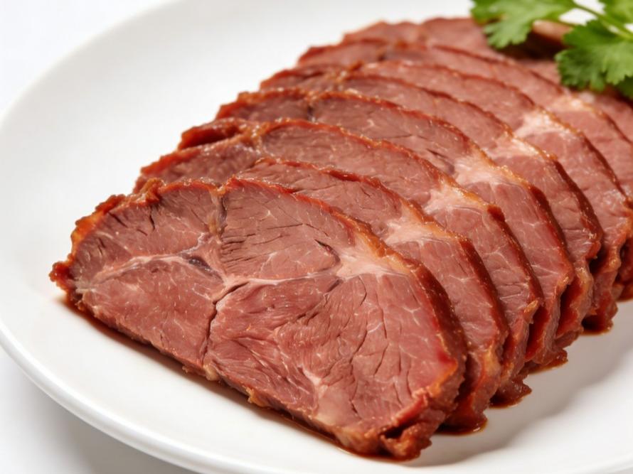
At this pivotal stage of plant-based meat products entering the mainstream market, a decisive consumer barrier is becoming increasingly apparent: color. Consumer expectations for plant-based meat have long surpassed the basic requirement of “looking like meat,” instead demanding “looking like real meat”—that vibrant, lifelike color transformation that naturally occurs during cooking. Currently, many products face the limitation of “static coloring”: a plant-based patty may retain a nearly uniform red hue before and after frying. This stands in stark contrast to real meat's dynamic transformation during heating—shifting from bright red to light brown while developing an enticing, glossy sheen.
This challenge stems from three closely intertwined technical pain points:
1. Absence of dynamic color transformation: The color evolution in real meat during cooking stems from the oxidation and denaturation of myoglobin under heat. Replicating this requires a pigment system capable not only of delivering the initial “raw meat red” but also undergoing controlled, progressive color shifts when heated—not merely fading or charring.
2. Textural Visual Distortion: Authentic meat coloration isn't a uniform coating. It features textured variations mimicking muscle fiber and fat distribution. Current coloring approaches often result in a “plasticine-like” homogeneous appearance, lacking visual depth and juiciness cues.
3. Stability Under Rigorous Processing: Plant-based meat production involves intense processing like high-moisture extrusion and high-temperature steaming. Subsequent frozen storage and home cooking (pan-frying, grilling, deep-frying) present ultimate tests for pigment heat resistance and migration resistance. Color must remain stable and authentic throughout, without fading, unevenness, or bleeding.
Therefore, solving plant-based meat's color challenge requires not merely finding a redder pigment, but a systematic, biomimetic, dynamic color solution capable of withstanding complex processing chains. This is precisely the target of Green Spring Technology's innovation centered on Paprika Oleoresin—we aim not only to make plant-based meat “look like meat,” but to make it “become meat” in the most authentic way possible.
Part One: The Bionic Coloring Principle of Capsicum Resin
The key to solving plant-based meat's color challenge lies in deeply understanding and mimicking the optical and chemical behavior of real meat. Green Spring Technology has built a coloring system based on bionic principles, centered around capsicum resin. Its scientific rigor is demonstrated through precise simulation across three levels.
I. Optical Bionics: Recreating Meat's “Optical Fingerprint”
Real meat's color originates from myoglobin's selective absorption and reflection of light. Our core technology enables chili oil resin to replicate this unique optical property within plant-based protein matrices.
· Precise spectral matching: Through refined processing, we optimize the spectral absorption curve of Paprika Oleoresin (primarily composed of capsanthin and capsanthin-like pigments). This ensures its reflectance characteristics at key wavelengths closely match those of myoglobin in specific states (e.g., fresh, medium-rare). This forms the physical foundation for achieving a “realistic appearance.”
· Three-dimensional hierarchical construction: We do not simply mix pigments uniformly. Using patented carrier and dispersion technology, we control the interaction between pigment particles and plant protein fibers. This ensures non-uniform distribution within the protein network during extrusion or restructuring. This microscopic unevenness manifests macroscopically as natural depth and fiber texture resembling muscle grain, eliminating the plastic appearance of “dyed modeling clay.”

II. Thermal Response Mechanism: Simulating Dynamic Color Change During Cooking
The true magic of cooking lies in dynamic color transformation. We developed a “Thermally Triggered Synergistic Color-Changing System.”
· Smart Base Color (Capsicum Resin): Provides a stable, vibrant initial “raw meat red” hue with excellent processing tolerance.
· Browning Trigger Component: Scientifically introduces trace amounts of heat-sensitive natural ingredients that undergo controlled reactions with capsanthin. Upon heating, this system activates precisely: the dominant carotenoid hue subtly shifts toward warm brown tones while the trigger component generates a Maillard-like browning undertone. Their synergy achieves a natural, progressive color transition from vivid red → pink → light brown—not abrupt changes or fading.
III. Gloss and Juiciness Suggestion: Evoking Visual Associations
A juicy piece of cooked meat possesses a specific surface sheen. We leverage the natural liposolubility of capsicum resin, combining it with carefully selected plant-based oil carriers.
· Lipid-Assisted Coloration: Pigments dissolved in lipids migrate and melt during cooking, creating a deep, naturally saturated color that permeates from within rather than sitting superficially.
· Gloss Modulation: When heated, fats form an ultra-thin film on surfaces that reflects light, creating a natural, oily sheen. This distinct luster—unlike simple water reflections or colloidal glare—sends a crucial visual signal to the brain, anticipating a “juicy,” “fat-rich” texture.
Through this integrated bionic system—spanning static spectral matching, dynamic thermal color change, and surface optical property control—chili oil resin transcends the singular coloring function of traditional pigments. It evolves into an intelligent color system that interacts with the cooking process, ultimately revealing the “cooked meat-like” vitality. In Part Three, we will explore how this scientific principle translates into concrete, mass-producible application solutions.
Part Two: Green Spring Solutions: The Complete Application Path from Lab to Production Line
Leveraging deep insights into plant-based meat color science, Green Spring Technology transforms the bionic coloring principle of chili oil resin into a systematic, modular solution. We provide not only core ingredients but also a complete application pathway—from formulation design to process adaptation—ensuring clients efficiently translate their ideal “cooked meat” color into stable, mass-produced quality.
I. Core Product System: Modular Color Building Blocks
To meet the diverse needs of different plant-based meat products, we have broken down our solution into three flexibly combinable functional modules:
1. Base Red Module (GR-PlantBase Red Series)
· Function: Provides a highly stable, colorimetrically precise initial “raw meat red” base tone. Utilizes protective technologies like microencapsulation to maintain activity during high-temperature, high-shear processing, serving as the foundation for all color builds.
2. Dynamic Browning Module (GR-PlantDynamic Brown Series)
· Function: Acts as a “heat-activated trigger.” Unlike traditional pigments, this module consists of specific natural thermosensitive components. When heated during cooking, it undergoes a controlled reaction with the Base Red module, synergistically producing progressive browning to precisely simulate the dynamic color spectrum from medium rare to well done.
3. Texture & Gloss Module (GR-PlantGloss & Texture Series)
· Function: This module is an oil-based carrier system preloaded with optimized pigments and flavors. It ensures perfect dissolution and dispersion of pigments within the fat phase. By regulating oil melting points and release characteristics, it creates a natural oily sheen and marbled fat distribution visual effect when the end product is heated, significantly enhancing juiciness and realism.
II. Key Process Adaptation Guide: Tailoring Color to Craft
We understand that coloring solutions must seamlessly integrate with production processes. Green Spring Technology provides validated integrated solutions tailored to mainstream plant-based meat production processes:
Production Process | Core Challenges | Green Spring Technology Adaptation Solution | Key Advantages |
High-Moisture Extrusion
| High temperature, high shear, high water activity—pigments are prone to degradation or loss. | Dry Mix Pre-Dispersion Solution: Base red and dynamic browning modules are uniformly premixed with plant protein powder in powder form before entering wet extrusion. | Colorants disperse uniformly within the protein network, resisting high-temperature shear. Color develops from the interior outward with exceptional stability. |
Emulsified Meat Products (e.g., Meat Patties, Meatballs)
| Requires uniform coloring within the fat emulsion system to prevent color bleeding (“bleeding” phenomenon).
| Oil-Phase Pre-Dissolution Solution: Pre-dissolve the Base Red and Texture-Gloss Module in the formulation's vegetable oil, then directly incorporate this oil phase into the emulsification process.
| Color is locked within fat globules, releasing as fats melt during cooking. This results in natural color penetration without bleeding, delivering excellent gloss. |
Re-engineered/Formed Products (e.g., Meat Chunks, Steaks) | Require simulation of complex muscle texture and surface caramelization layers. | Staged Coloring and Surface Treatment Solution: Internal application via dry mixing or injection of base red; surface application via spraying or immersion in a solution containing a dynamic browning module, forming a realistic “crisp brown layer” during subsequent cooking. | Achieves three-dimensional color and texture, with dynamic and lifelike surface color reactions, delivering extremely high biomimetic realism. |
III. Integrated Services: From Concept to Shelf
We offer more than just product portfolios—we provide end-to-end services:
· Rapid Formula Matching: Based on your protein source (pea, soy, wheat, etc.), processing method, and target product form (mince, chunks, slices), we recommend optimal module combinations and starting addition levels.
· Pilot and Scale-Up Support: Assist you through the entire process—from sample production to process parameter optimization—in our lab and pilot facilities.
· Stability & Compliance Documentation: Provide comprehensive processing stability data, shelf-life projections, and regulatory support documents meeting clean label requirements.
Through this complete pathway of “Modular Products + Process Customization + Full-Cycle Service,” Green Spring Technology makes achieving authentic “cooked meat-like” color clear, reliable, and efficient. In the next section, we'll demonstrate the real commercial value this solution delivers with actual data and case studies.
Part Three: Green Spring Technology Support
I. Professional Service System
1. Free Samples & Diagnosis: Provide dedicated plant-based meat coloring sample packs and conduct color optimization diagnostics for your existing products.
2. Custom Development: Tailor exclusive color solutions based on your protein base, process, and brand positioning.
3. Compliance & Mass Production Assurance: Solutions meet clean label requirements with end-to-end quality tracking from pilot production to large-scale supply.
II. Get Your Solution Now
1. Submit your needs to receive free samples and the Plant-Based Meat Color Application Guide
2. Schedule a technical meeting to discuss customized solutions
3. Contact us for the latest case study data
Green Spring Technology Plant-Based Food Color Research Center
Hotline: +86 13649243917
Email: helen@greenspringbio.com
Official Page: https://www.greenspringnatural.com/
Infusing plant-based meat with authentic vitality through scientific coloring.
-
Prev
Breakthrough Application of Paprika Oleoresin in the Beauty and Cosmetics Sector
-
Next
The Definitive Guide to Paprika Oleoresin: Compliance, Regulations, and Clean Label Strategies