How to Synthesize Ginkgo Extract Ginkgolic Acid?
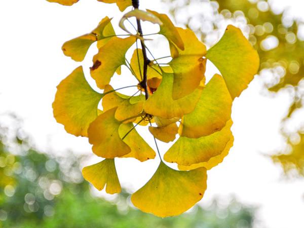
Ginkgo biloba L. is the oldest living tree species on the planet and is known as the “golden living fossil”. As a unique economic gymnosperm in China, it has multiple values as food (white fruit), medicine (leaves and white fruit) and ornamental plants. Ginkgo nuts (seeds) are a famous health-promoting dried fruit in China. As a food with medicinal properties with high nutritional value, the ginkgolides contained in it are potential active ingredients against the 2019-nCoV virus[1]. Ginkgo biloba leaves are rich in flavonoids and lactones. Extraction of Ginkgo biloba (EGB) with acetone-water as the extracting agent can further enrich the two active substances to effectively treat cardiovascular and cerebrovascular diseases [2]. Ginkgo biloba is a world-famous ornamental tree for landscaping with its unique leaf shape, golden leaf color and upright tree shape. At present, ginkgo has been extensively developed in the pharmaceutical, food, cosmetics, bonsai and timber industries, and is one of the key economic tree species for the current and future revitalization of the rural economy and the construction of a beautiful China.
Since the 1970s, China's ginkgo resources have grown rapidly, and the existing resources account for more than 80% of the world's total. Ginkgo trees are distributed in all provinces (including those introduced from other places), except for Hainan, Heilongjiang and Inner Mongolia. At present, the cultivated area of ginkgo in China is more than 333,300 hm2, with an annual output of about 11,000 tons of ginkgo nuts and 10,000 tons of dried green leaves. The annual output value of the primary industry of ginkgo cultivation is about 2 billion yuan, and the secondary industry of ginkgo processing and pharmaceutical production (ginkgo leaf medicine) is worth more than 10 billion yuan.

However, about 30,000 tons of outer seed coats are discarded each year due to production. Zhang Xinhui et al. [3] showed that the outer seed coat contains active substances such as ginkgolic acid, flavonoids, terpene lactones and polysaccharides. Itokawa et al. [4] showed that ginkgolic acid is another important active substance in ginkgo biloba, in addition to ginkgolide and ginkgoflavone. It has bactericidal, bacteriostatic, anti-inflammatory, anti-viral, insect repellent and insecticidal effects, and is of great value for development. Therefore, the large amount of discarded outer seed coats not only pollutes the environment but also causes a waste of resources.
In the 1970s, Gellerman et al. [5–7] detected the phenolic acid ginkgolide from ginkgo leaves, immature seeds and mature outer seed coats. Subsequent quantitative measurements showed that ginkgolide is concentrated in mature outer seed coats. Wang Jie et al. [8] and Li Hongqing et al. [9] used mass spectrometry to identify four ginkgolic acids and two ginkgolides from ginkgo acidic extracts. Modern medical research has proved that ginkgolides have certain allergenic toxicity and cytotoxicity, can resist the growth of bacteria and fungi, and have the effect of killing pests.
Field cultivation experiments have proved that ginkgolides can be developed as biopesticides, and they may be used in a wide range of fields such as medicine and cosmetics in the future. However, compared to substances with clear pharmacological activities such as ginkgo flavonoids and terpene lactones, the biosynthesis pathway, regulatory mechanism and derivative efficacy of ginkgolic acid have not been studied in depth, which has slowed down the research and development of molecular biology techniques to inhibit the synthesis of ginkgolic acid and the engineering application of fermenting to produce high yields of ginkgolic acid, thus hindering the further development of the ginkgo industry.
In view of this, the author summarizes the basic research on the biological activity of ginkgolides in recent years, details the synthesis pathway of ginkgolides, which is composed of fatty acid synthesis and polyketide synthesis, summarizes the catalytic mechanism of the key enzymes involved in the synthesis, and looks ahead to the future research direction, with the aim of providing reference and theoretical support for subsequent research on ginkgolides.
1 Biological activity of ginkgolides
Ginkgolide acid belongs to the phenolic lipid family, which has four main categories: alkylphenols, alkylresorcinols, anacardic acids and alkylcatechols [10]. They are a class of secondary metabolites in plants. Different species of plants may contain unique phenolic substances. Their main physiological function is to resist biotic and abiotic stresses.
1.1 Ginkgolide composition and physical and chemical properties
Ginkgolides are important secondary metabolites of ginkgo biloba, and belong to the class of lacquers. They include three components: ginkgolic acid, ginkgol and bilobol [11]. Ginkgolic acid is a 2-hydroxy-6-alkenyl (alkynyl) benzoic acid (Fig. 1a) with a side chain length of 13, 15, or 17 and a side chain double bond number of 0 to 2. A total of five types have been isolated and identified, namely, ginkgolic acid B, ginkgolic acid A, ginkgolic acid, heptadec-1-enylginkgolic acid and heptadec-1-dienylginkgolic acid. Ginkgolides are 3-alkenylphenols with a side chain length of 15 or 17 and a side chain double bond count of 1, and there are two types in total; ginkgolic acids are 5-alkenylresorcinols with a side chain length of 15 or 17 and a side chain double bond count of 2 (Fig. 1b), and there are two types in total.
Plants such as geranium (Pelargonium hortorum) in the Geraniaceae family [12] and sumac (Anacardium occidentale) in the Anacardiaceae family [13] also contain urushiolic acids. The alkyl/alkenyl side chain lengths and the number of unsaturated bonds, and so on, but the chemical structure is similar to that of ginkgolides. Ginkgolic acids are the main substances in ginkgolides extract, accounting for 90% of the entire acidic extract. The proportions of the five ginkgolic acids are different, mainly including ginkgolic acid (50% content), followed by heptadec-1-enyl ginkgolic acid (22%) and ginkgolic acid (20%) [14] (Figure 2). With the development of science and technology, ginkgo acid has been detected in ginkgo leaves, outer seed coats and kernels. The total ginkgo acid content of ginkgo leaves from different varieties (strains) is about 14.5765-23.6813 mg/g [15], and the total ginkgo acid content of mature kernels is about 0.11 mg/g [14]. and the total ginkgolic acid content in the outer seed coat can reach 28.78 mg/g [16].
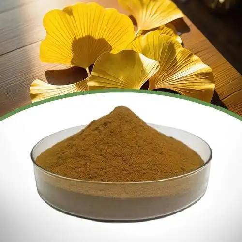
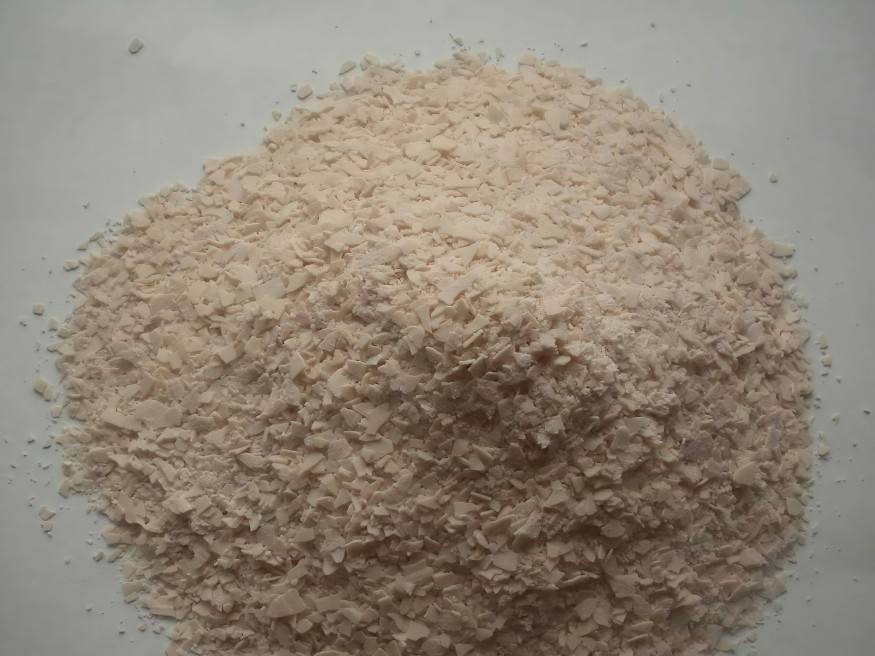
The melting point of ginkgolic acid is 41-42°C. At room temperature, pure ginkgolic acid is oily or powdery in appearance, and is poorly soluble in polar solvents such as water or ethanol, but easily soluble in non-polar solvents such as light petroleum ether. It precipitates as crystals in saturated petroleum ether solvent [17]. In solution, the hydroxyl and carboxyl groups on the phenolic acid benzene ring ionize to produce a weak acid, which can undergo esterification and saponification reactions. Ginkgolic acid that has undergone esterification or saponification is easier to extract and separate, achieving the effect of purification. The melting point of ginkgolic acid is about 136 °C and the boiling point is about 500 °C. At 200 °C, the carboxyl group on the phenolic acid benzene ring will undergo a decarboxylation reaction to release CO2, which can be separated using the temperature gradient method. Therefore, according to physical and chemical property research, in the processing of ginkgo products, methods such as “hot air cooking”, “ultrasonic-assisted extraction of ginkgo phenolic acid with resin adsorption” and “compatibility of Chinese medicinal herbs” are often used to remove phenolic acids to achieve the purpose of detoxification. Chen et al. [18] latest research results show that the laccase immobilized on a new type of electrospun nanofiber felt can catalyze the degradation of ginkgo phenolic acid. However, the above-mentioned methods for dephenolic acid treatment have disadvantages such as high cost and difficulty of operation, and are not suitable for large-scale detoxification.
1.2 Biological activity of ginkgolides
Ginkgolides have antibacterial, insecticidal, allergenic, cytotoxic and anticancer effects. Their diverse biological activities can be exploited in agricultural production, healthcare and other fields.
1.2.1 Antibacterial effect
Ginkgo acid is 6-alkylsalicylic acid, which has a wide range of antibacterial properties. It has an inhibitory effect on Bacillus subtilis, Escherichia coli, Staphylococcus aureus, Shigella dysenteriae, Pseudomonas aeruginosa, and a variety of Gram-negative and Gram-positive bacteria [19-20]. Its acidic extractant has an inhibitory effect on the rice sheath blight fungus, the tomato wilt fungus, the apple anthracnose fungus, the corn leaf spot fungus and the red mold fungus, and its antibacterial effect is comparable to that of some antifungal drugs [21]. Xu Lichun et al. [22] showed that 0.1% ginkgoic acid (15:1) had an effective rate of 92% in inhibiting fungi, while 0.5% clotrimazole had only 68% effectiveness. Muroi et al. [23] showed that ginkgoic acid had a synergistic effect with standard antibacterial drugs in killing methicillin-resistant Staphylococcus aureus, and the bactericidal activity of the combination was at least 100 times higher than that of the single drug within 48 hours.
1.2.2 Insecticidal effect
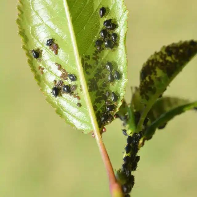
Ginkgolide acid has been shown to have a significant killing effect on aphids, grubs, cabbage moths, spider mites, mulberry ticks, rice borer and other chewing mouthparts insects [20]. In a test to control aphids, Shi Qitian [24] found that the killing effect of ginkgolide acid extract was comparable to that of the pesticide imidacloprid. Deng Yecheng et al. [25] used different polarities of exocarp extracts in contact toxicity tests and found that they had a strong killing effect on the brown planthopper, peach aphid, red spider and cabbage butterfly larvae.
1.2.3 Allergic effects
In 1934, Hill et al. [26] reported that an active substance in ginkgo has a strong erosive effect on the skin. In 1969, Gellermen isolated lacquer phenolic acids from cashew nuts and ginkgo seeds, respectively, and pointed out that they have a strong sensitizing effect on the skin [5]. Cheng Liang et al. [27] reported that ginkgolic acid (C15:1) is metabolized to ginkgolide, which is further oxidized to form catechol, causing an allergic reaction. Vincieri et al. [28] showed that ginkgolic acid can inhibit the activity of various dehydrogenases in glucose metabolism. Ahlemeyer et al. [29] found that ginkgo acid has a competitive inhibitory effect on glycerol-3-phosphate dehydrogenase. Some scholars speculate that ginkgolic acid has a bipolar nature (hydrophilic and lipophilic) and can inhibit the activity of enzymes related to the body or organs, thereby affecting metabolism and causing allergies [30].
1.2.4 Cytotoxicity
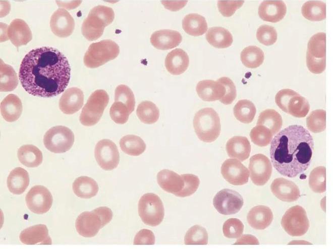
Ginkgolide has both hydrophilic and lipophilic groups, which can bind to the cell membrane across the membrane and cause cell death. Al-Yahya et al. [31] used ginkgo extract containing ginkgo acid to conduct a toxicity test on male rats, and the results showed that the extract can cause the loss of chromosomes such as germ cells, affecting the normal reproductive function of rats. Ahlemeyer et al. [29] found that ginkgolide acid has neurotoxicity and can lead to the death of chick neuroblastoma cells. In recent years, researchers have conducted separate studies on the toxicity of various ginkgo acids. Jiang et al. [32] found that ginkgo acid (C15:1) can cause oxidative stress and damage to rat liver cells by inducing purine metabolic disorders. Yao et al. [33] found that the toxicity of heptadecylidene ginkgo acid (C17:1) to HepG2 cells is directly proportional to time and dose, and enhanced cytotoxicity by mediating CYP1A and CYP3A metabolism. Studies have shown that among the ginkgolides, ginkgolic acid (C13:0), ginkgolic acid (C15:1) and heptadecylidene ginkgo acid (C17:1) are more toxic, and other phenolic acids can act in combination with the three ginkgo acids to increase cytotoxicity.
1.2.5 Anticancer effect
Ginkgolides have cytotoxicity and can play an anticancer role. Qiao et al. [34] found that ginkgo acid can induce the activation of adenosine monophosphate-activated protein kinase (AMPK) to inhibit the proliferation and migration. Liang et al. [35] found that ginkgo acid can inhibit the growth of gastric cancer cells by inhibiting the ROS-regulated STAT3/JAK2 signaling pathway. Liu et al. [36] showed that ginkgo acid can block the transition of colon cancer cells from the G0/G1 phase of cell division, leading to cancer cell death. In vitro cell experiments have shown that ginkgo acid can inhibit the proliferation and migration of cancer cells and activate related enzymes to promote apoptosis. and may become an adjuvant anti-cancer drug for tumor treatment in the future.
2 Ginkgolide biosynthesis
In the 1970s, Gellerman from the University of Minnesota in the United States used the 14C labeling method to conduct a preliminary exploration of the ginkgolide acid synthesis pathway. In the 1990s, Walters et al. [37] used gas-liquid chromatography (GLC trapping) to analyze labeled monomethyl esters extracted from liquid chromatography (HPLC), and determined the specific synthetic steps of urushiol (2-hydroxy-6-alk(en)yl-benzoic acid) in geranium. Singhal [38] and Narnoliya et al. [39] further investigated the types and functions of key enzymes in the synthesis of urushiol.
2.1 Ginkgolide biosynthetic pathway
Gellerman et al. [6–7] believe that the aromatic ring and long-chain alkyl/alkenyl group of ginkgolide are synthesized stepwise, According to experimental inference, it is divided into three parts: ① malonyl-CoA and acetyl-CoA form free palmitoyl-CoA and oleoyl-CoA through fatty acid synthesis; ② long-chain acyl-CoA is synthesized to form ginkgolic acid through polyketide synthesis; ③ ginkgolic acid loses the carboxyl group of the benzene ring and is oxidized and reduced to substances such as ginkgol.
Geranium (Pelargonium hortorum) is a plant in the Geraniaceae family. Its trichomes only secrete shikimic acid (a homolog of ginkgoic acid), making it the best species for studying the shikimic acid synthesis pathway. Wild-type geranium secretes shikimic acid with a saturated side chain, while insect-resistant types secrete shikimic acid with a monounsaturated side chain (C15:1 and C17:1). Studies have found that the unsaturated urushiol acid in geraniums is identical in molecular structure to the two ginkgolic acids (ginkgolic acid (C15:1) and ginkgolic acid (C17:1)) except for the difference in the position of the double bond. Hesk et al. [12] compared the differential genes and metabolites of wild-type and insect-resistant geraniums, and revealed the molecular mechanism of urushiol synthesis. The researchers used RNA-seq technology to construct a geranium cDNA transcriptome database, and identified the key genes for synthesis to be the Polyketide Synthases Ⅲ (PKS Ⅲ) gene and the Stearyl-ACP Desaturase (SAD) gene through gene annotation comparison and differential expression analysis. The regulatory pathway of ginkgolide synthesis is explained using ginkgolic acid (15:1) and heptadecylideneginkgolic acid (17:1) as examples.
2.1.1 Synthesis of the alkyl side chain
The synthesis of ginkgolides is based first on the synthesis of palmitoleic acid and oleic acid, i.e. the synthesis of the alkyl side chain (Figure 3). The fermentation of stored sucrose into phosphoenolpyruvate, the allosteric change of pyruvate kinase (Pyruvate kinase) to form pyruvate from the cytoplasm into the plastid, and the catalysis of pyruvate dehydrogenase (Pyruvate Dehydrogenase, PDH) to form acetyl-CoA provide the starting 2C molecule for fatty acids. Acetyl-CoA is catalyzed by acetyl-CoA carboxylase (Ace- tyl-CoA Carboxylase, ACC) to form malonyl-CoA. Transacylase (TA) replaces the CoA molecule with an acyl carrier protein (ACP) to form malonyl-ACP. Malonyl-ACP loses its carboxyl group to become acetyl-ACP, and is condensed in a cycle by β-Ketoacyl-ACP synthase III (KAS III) to form the starting material 4C acyl-ACP.
4C acyl-ACP is repeatedly condensed by β-Ketoacyl-ACP synthase I (KAS I) to form palmitoyl-ACP (C16:0 ACP), which is then dehydrogenated by Δ9-stearoyl-ACP desaturase (SAD) to form palmitoleic-ACP (Δ9 C16:1 ACP). SAD) is dehydrogenated to palmitoleic acid-ACP (Δ9 C16:1 ACP), and then catalyzed by ketoacyl synthase II (β-Ketoacyl-ACP synthase Ⅱ, KASⅡ) to form oleic acid-ACP (Δ11 C18:1 ACP). Palmitoyl-ACP and oleoyl-ACP in the plastids are respectively deacylated by thioesterase A (TE/FatA) and thioesterase B (TE/FatB) to form free unsaturated fatty acids [40]. Finally, the free fatty acids are converted into palmitoleyl-CoA (Δ9 C16:1CoA) and oleoyl-CoA (Δ11 C18:1CoA) by acyl-CoA synthase (ACS) in the outer membrane of the plastid, and transferred to the cytoplasm to become the two important precursors of ginkgolic acids.
However, the fatty acid synthesis pathways of ginkgolic acid and geranylgeranyl are different. The precursors for the synthesis of ginkgolic acid (C15:1) and heptadecylideneginkolic acid (C17:1) are palmitoleic acid (Δ9 C16:1) and oleic acid (Δ11 C18:1), while the precursors for the synthesis of the 2 monoenyl side chains of oleic acid are palmitoleic acid (Δ11 C16∶1) and oleic acid (Δ13 C18∶1). The common ω-7 fatty acids (palmitoleic acid (Δ9 C16∶1) and oleic acid (Δ11 C18∶1)) can be formed in many wild plants and algae from palmitoyl-ACP (C16∶0 ACP) by desaturation, polyketone and other reactions. Schultz et al. [41] found that a novel type of geranyl ACP desaturase in geranium dehydrogenizes myristic acid-ACP (C14:0 ACP) to form myristic acid-ACP (Δ9 C14:1), and then undergo polyketide and other reactions to form two monounsaturated fatty acids (palmitoleic acid (Δ11 C16:1) and oleic acid (Δ13 C18:1)). Therefore, the synthetic pathway of the precursor fatty acids of ginkgolic acid is easier to explore than that of pelargonolic acid in geraniums.
Singhal [38] identified and verified the expression of genes related to the synthesis of monounsaturated fatty acids (palmitoleic acid and oleic acid) in geranium. The results showed that when the gene expression of acyl carrier proteins (ACPs), ketoacyl synthases (KASs) and thioesterases (TEs) in the tissue was high, the content of fatty acids and ursolic acid was also high. Acyl carrier protein (ACP) is a conserved intermediate carrier that is central to the fatty acid synthesis process. It works with acyl-ACP desaturase (AAD) to desaturate the acyl chain, changing the ratio of saturated and unsaturated fatty acids. It can also act as a rate-limiting enzyme together with ketoacyl-ACP synthase (KAS) to regulate the rate of phenolic acid synthesis.
2.1.2 Phytylbenzene ring synthesis
Phytylbenzene ring synthesis is a key step in the formation of phenolic acids from higher fatty acids. The synthesis pathway is shown in Figure 4. Taking gammacerolic acid (C15:1) as an example, first, palmitoyl (Δ9 C16:1) from the fatty acid pathway uses malonyl-CoA as a substrate and under the action of Polyketide Syn- thase Ⅲ (PKSⅢ), four carbon atoms are added to the acyl end through a two-step condensation reaction; second, the ketone group at the C3 position is reduced by the ketone reductase (PKS- ketoacyl-CoA reductase, KR) to form a hydroxyl group, and a double bond is formed by the dehydratase (PKS-dehydratase, DH) removes a molecule of water to form a double bond; third, after condensation and polymerization, two more carbon atoms are added to the acyl end. At this time, the hydrogen ion at the C2 position moves closer to the ketone group at the C7 position to maintain the stability of the 4,15-diene tripeptide intermediate structure; fourth, ensuring that the ketone group at C1 does not decarboxylate, the cyclase (PKS-Cyclase) catalyzes the cyclization of the hydrogen ion at C2 with the ketone group at C7, similar to an aldol condensation, and a double bond is formed under the action of the dehydratase (PKS-dehydratase); fifth, the enoyl reductase (PKS-enoyl reductase, ER) catalyzes the formation of a double bond and aromatization of the ketone group on the hexagonal carbon ring. It and the C1 carboxyl group together form the benzoic acid structure; finally, the monounsaturated side chain is formed to produce ursolic acid (C15:1) [37, 39].
Plant type III polyketide synthase (PKS III) is the rate-limiting enzyme for the synthesis of phenolic lipids (alkylphenols, alkylresor- cyls, urushiols, alkylcatechols, etc.) [42]. Chalcone synthase (CHS) and stilbene synthase (STS) are two of the most representative superfamilies. STS) are two of the most representative superfamilies. They can catalyze the condensation and cyclization of acyl chains at the C1 and C6 positions (Claisen condensation) and at the C2 and C7 positions (Aldol condensation), respectively, to form phenolic substances. However, uronic acids can catalyze the condensation of the hydrogen ion at the C2 position with the ketone group at the C7 position while retaining the carboxyl group at the C1 position to form benzoic acid. This polyketide cyclization reaction that retains the benzene ring carboxyl group is relatively rare in plants. Therefore, further research on polyketide synthase type III (PKS III), cyclase (PKS-Cyclase) and ketoacyl reductase (PKS-ketoacyl-COA reductase, KR) can further reveal the mechanism of ginkgolic acid synthesis, and the content of phenolic acids in ginkgo tissues can be controlled by combining physical and chemical or biotechnological methods.
2.2 Key enzyme genes for phenolic acid synthesis
At present, the main reports on the research of key enzyme genes for phenolic acid synthesis come from urushiol, while there are few reports on ginkgolides. The phenolic acid synthesis pathway is divided into 2 parts: fatty acid synthesis and aromatic ring synthesis. Among these, acyl carrier protein (ACP), stearoyl desaturase (SAD), ketoacyl-ACP synthase (KASs), type III polyketide synthase (PKS III), and cyclase (PKS-Cyclase) are key enzymes that determine the rate and content ratio of urushiol synthesis.
2.2.1 Acyl carrier protein (ACP)
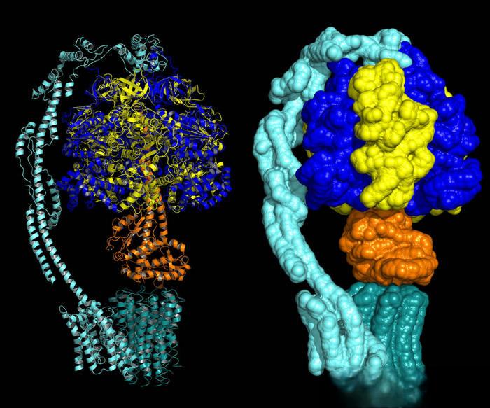
Acyl carrier protein (ACP) belongs to the large family of carrier proteins. As a mixed protein, it can associate with various protein-enzyme complexes to transfer acyl chains from one enzyme center site to another. It also acts as a cofactor for stearoyl-ACP desaturase (SAD) and acyl-ACP hydrolase (AAH), and plays an important role in the fatty acid synthesis (FAS) and polyketide synthesis (PKS) pathways.
Li Mengjun et al. [43] analyzed the structure of acyl carrier protein genes in 17 species, including Arabidopsis thaliana, Clycine max, Oryza sativa and Zea mays, and classified them into five categories. Among them, the plasmid-type ACP gene family (the coding region consists of 4 exons and 3 introns) and the mitochondrial-type ACP gene family (the coding region consists of 2 exons and 1 intron) account for the largest proportion of the whole. They both have a very conserved serine site that can bind to the 4'-phosphopantetheine adduct cofactor group, and activate the holo-ACP to function. The tertiary structure of the ACP protein family is mostly consistent and consists of four conserved α helices. Three of the α helices (I, II, IV) are parallel to each other, while the α helix (III) is perpendicular to the helix center, forming a hydrophobic structural cavity providing a protective hydrophobic structure for various acyl chains [44].
The α helix Ⅱ can interact with ketoacyl-ACP synthase Ⅱ (β-Ketoacyl-ACP synthase Ⅱ, KAS Ⅱ) to further extend the acyl chain, and is known as the recognition helix. Singhal [38] performed transcriptome sequencing on geraniums and screened out two complete ACP protein cDNA sequences (Pxh1 and Pxh2) related to urushiol acid synthesis. Gene expression verification and phylogenetic analysis showed that the Pxh1 and Pxh2 genes were highly expressed in the trichome tissue of geranium, and are highly homologous to the 14-carbon-1-ene acid (Δ9 C14:1) biosynthesis ACP protein gene of coriander (Coriandrum sativum), which catalyzes the desaturation of fatty acids in the uronic acid pathway.
2.2.2 Ketoacyl-ACP synthase (KASs)
Under the action of the fatty acid synthase (FAS) complex, malonyl-ACP is used as a substrate to form the precursors of ginkgolic acid, palmitoleic acid (Δ9 C16:1) and oleic acid (Δ11 C18:1), through multiple condensation cycles. The fatty acid synthase complex is composed of three ketoacyl-ACP synthases (KASs), which belong to the superfamily of cond-enzymes. They are all hydrophobic lipoproteins containing the KAS conserved structural domain and no signal peptide. KAS III catalyzes the polymerization of malonyl-CoA and acetyl-CoA to form the initial acyl chain (3-ketobutyryl-ACP); KAS I catalyzes the polymerization of the acyl chain and malonyl-ACP to form fatty acids with 6C-16C; and KAS II catalyzes the condensation of malonyl-ACP and palmitic acid to form 18C fatty acids.
In plants such as perilla [45] and upland cotton [46], KAS II is a slightly acidic non-secreted protein with an amino acid length of about 500 aa. It determines the ratio of C16:C18 fatty acids and can regulate plant cold resistance. Researchers have obtained seven ketoacyl-ACP synthase (KASs) genes from the transcriptome of Pelargonium xanthium, of which PxKASⅠa, PxKASⅠb and PxKASⅠc have high and stable expression in the trichome tissue of Pelargonium xanthium, while the gene expression in other tissues is low. Further treatment of trichomes with temperature gradients (18, 23 and 28 °C) showed that the expression of the three genes decreased with increasing temperature, and was positively correlated with the content of fatty acids (palmitoleic acid (Δ11 C16:1) and oleic acid (Δ13 C18:1)) and urushiol, or involved in the urushiol synthesis pathway. Evolutionary analysis shows that the KAS gene family of geranium is highly homologous to the ketoacyl synthase (KAS) genes of Arabidopsis thailana and Glycine Max, and the related catalytic mechanism is relatively clear. However, there have been few molecular studies on the KAS of ginkgolide ketoacyl-ACP synthase (KAS). how the precursor palmitoleic acid (Δ9 C16:1) and oleic acid (Δ11 C18:1) of ginkgo acid (C15:1) and heptadecenoic acid (C17:1) are regulated by the ginkgo ketoacyl-ACP synthase family as important monoenriched unsaturated fatty acids remains to be explored in depth.
2.2.3 Stearoyl-ACP desaturase (SAD)
Stearoyl-ACP desaturase (SAD) is the only known family of soluble desaturases in plants, which regulates the ratio of saturated to unsaturated fatty acids. Among them, Δ9 stearoyl-ACP desaturase is the most widely studied in plants. It catalyzes the dehydrogenation of the C9 and C10 positions of the acyl chain to form the first double bond. The SAD protein is a homodimer composed of a conserved domain belonging to the acyl-ACP desaturase family and a conserved domain belonging to the ferritin family. The four α helices in the SAD conserved region form a four-helix bundle structure, which conceals a symmetrical Fe-O-Fe two-iron cluster catalytic center. Together, they form the active center of the enzyme for acyl chain dehydrogenation [47]. Protein crystal structure shows that SAD enzyme has a deep groove extending from the molecular surface to the interior, which can accommodate the 18C acyl chain and bind to the ferrous center at the bottom of the groove to undergo redox reactions. This groove structure can also accommodate 16C and 14C acyl chains to react, but the catalytic turnover rate is low [48].
Schultz et al. [41] screened a novel acyl-ACP desaturase from a geranium cDNA library. Gene sequence alignment revealed a high degree of homology with the castor bean stearoyl-ACP desaturase gene (SAD). The results of genetic transformation and verification using Escherichia coli (E. coli) showed that the novel gene can catalyze the dehydrogenation of myristic acid-ACP (C14:0) to form myristic acid-ACP (Δ9 C14:1), so it is named myristic acid-ACP desaturase gene (MAD, Δ9 14:0-ACP desaturase). Singhal [38] gene expression detection and tobacco (Nicotiana tabacum) genetic transformation validation results show that tetradecyl-ACP (Δ9 C14:1), the product catalyzed by myristic acid-ACP desaturase (MAD), is a key precursor of palmitoleic-ACP (Δ11 C16:1) and oleic-ACP (Δ13 C18:1). The activity of myristoyl-ACP desaturase (MAD, Δ9 14 ∶0- ACP desaturase) determines the content of palmitoyl-ACP ( Δ11 C16∶1) and oleoyl-ACP (Δ13 C18∶1) by detecting enzyme activity, fatty acid content and phenolic acid content, which in turn regulates the content of the two monoenyl side chains, oleic acid (C22:1 and C24:1). In addition, Singhal [38] set up a temperature gradient (18, 23 and 28 °C) to investigate the expression changes of related enzyme genes in geranium trichome tissue, and found that the expression levels of the myristic acid-ACP desaturase gene (MAD, Δ9 14 ∶0- ACP desaturase) and stearoyl-ACP desaturase gene (SAD, Δ9 18 ∶ 0-ACP desaturase) expression decreased with increasing temperature.
Wang et al. [49] cloned a stearoyl-ACP desaturase gene (SAD, Δ9 18:0-ACP desaturase) from Ginkgo biloba leaf cDNA and subjected Ginkgo biloba leaves to temperature stress (4, 15 and 45 °C). The results showed that the gene expression was high at low temperatures (4 °C) and room temperature (15 °C), while the gene expression at high temperatures (45 ℃) was several times lower than that of the control group. Subsequently, Liu Xinliang et al. [50] showed that the GbSAD gene encodes a peptide chain with a chain length of 412 aa and a molecular weight of 47 kDa. Cluster analysis showed a high degree of similarity with the stearoyl-ACP desaturase (SAD) amino acid sequences of other gymnosperms. Exogenous hormone experiments showed that the expression of the GbSAD gene was not regulated by abscisic acid (ABA), methyl jasmonate (MeJA) or ethylene (ETH), but salicylic acid (SA) activated gene expression, with the highest expression value being 9.7 times that of the control group, indicating that SA may be involved in the regulation of the fatty acid synthesis pathway.
Pelargonium graveolens and Ginkgo biloba contain the unique myristic acid-ACP desaturase (MAD, Δ9 14 ∶0-ACP desaturase) and stearic acid-ACP desaturase (GbSAD, Δ9 18 ∶0-ACP desaturase) respectively. Both enzymes are temperature-sensitive and highly active at low temperatures. Both enzymes are the only water-soluble desaturases in plants and and have a high degree of similarity in terms of their functional structure. Therefore, homologous function verification of the ginkgo stearoyl-ACP desaturase (GbSAD, Δ9 18:0-ACP desaturase) will further clarify the mechanism of ginkgolide acid synthesis.
2.2.4 Type III polyketide synthase (PKS III)
Polyketide synthases are divided into three categories: ①PKS I, known as modular PKS, is composed of several multifunctional polypeptides, each of which carries a unique, non-reusable catalytic domain. PKS II, also known as iterative or aromatic PKS, is an iterative system of a multi-enzyme complex that uses a set of reusable domains to catalyze the formation of phenol polyketone structures multiple times during repeated reaction steps. ③PKS III, known as the chalcone synthase-type enzyme, is completely different from the first two types of PKS enzyme families. It can reuse homologous bifunctional proteins, does not rely on the activation of ACP and its active site 4'-phosphopantetheine sulfide, and does not need to activate the acyl-CoA substrate through ACP to directly react with acyl-CoA. Although the structural mechanisms of different types of PKS are different, they all use the ketosynthase (KS) domain or subunit to catalyze the formation of C-C bonds, which decarboxylate and condense acyl-CoA to extend the carbon chain.
The PKS III family is very diverse. Chalcone synthase (CHS) and stilbene synthase (STS) are the two earliest discovered and most representative families. Their amino acid sequences are 60% to 75% similar [42]. The chalcone synthase (CHS) family is widely found in plants. It is a homodimeric protein with a molecular weight of 40–45 kDa. Using a highly conserved active center (a combination of Cys-His-Asn), it catalyzes the carbon chain elongation of coumarin CoA using three pyruvate-CoA substrates to form a tetrapeptide intermediate ring structure [42, 51–52]. The C6 hydrogen ion of the tetrapeptide intermediate ring undergoes a Claisen condensation reaction with the C1 ketone group to form a carbon hexa-membered ring, which is then aromatized to form a diphenyl ketone, also known as a chalcon (Figure 5). Chalcone is an important precursor for the synthesis of flavonoids. The stilbene synthase (STS) enzyme has been reported in plants and microorganisms (e.g. Streptomyces, yeast and bacteria). It has a molecular weight of about 43 kDa, is a dimer consisting of 2 subunits, and has a protein with a specific conserved domain IPNS(F) AGAIAGN, which is highly homologous to the chalcone synthase (CHS) [53].
The STS enzyme uses coumaroyl CoA as a substrate to form a tetrapeptide intermediate ring structure. The C7 ketone group of the tetrapeptide intermediate ring undergoes an Aldol condensation reaction with the C2 hydrogen ion to form a carbon six-membered ring, and then aromatized to form the phytoalexin resveratrol glycoside [54] (Figure 5). Singhal [38] identified a type 2 ketoacyl CoA synthase (KCS2, Keto-acyl CoA synthase 2) in geranium (Pelargonium hortorum), its tertiary spatial structure is similar to that of polyketide synthase type III (PKS III), and it is speculated to be involved in the cyclization condensation reaction in the urushiol synthesis pathway. Ketoacyl CoA synthase (KCS) is a rate-limiting enzyme that catalyzes the first condensation reaction in the synthesis of very long chain fatty acids (VLCFAs). Research on its gene family has mainly focused on the model plant Arabidopsis thaliana, and there are few reports of research in other plants. Costaglioli et al. [55] classified the 21 KCS gene members in Arabidopsis into four subgroups (FAE1, KCS1, FDH and CER6) based on gene homology and genetic evolution analysis. Among them, the FAE1 gene was the first KCS family gene cloned by James et al. [56] using the transposon tagging method. The amino acid sequence of FAE1 is highly homologous to other polyketide synthases (chalcone synthase (CHS), squalene synthase (STS), and type III ketoacyl-ACP synthase (KAS III)).
Ketol-acyl CoA synthase (KCS) from different species has different substrate specificities. Ginkgolide A belongs to the lacquer acid substances and its pathway-specific type III polyketide synthase (PKS III) is similar in terms of its functional structure to geranium's type 2 keto-acyl CoA synthase (Keto-acyl CoA synthase 2). In addition, the polyketide synthase of the urushiol acid pathway and the acyltransferase (STS) both recognize the C7 ketone group in the tetrapeptide intermediate ring and the C2 hydrogen ion to undergo an aldol condensation reaction to form a carbon hexa-membered ring. The difference is that the acyltransferase (STS) undergoes a decarboxylation reaction during the aldol condensation cyclization reaction, which oxidizes the ketone group at the C1 position to release carbon dioxide, However, during the polyketide cyclization of lactonic acid, the C1 ketone group is retained, and the benzoic acid structure is formed by the replacement of CoA with a hydrogen ion (H+). The current catalytic mechanism is not yet clear and requires further research.
2.2.5 Polyketide cyclase (PKS-cyclase)
2,4-dihydroxy-6-pentylbenzoic acid (Olivetolic acid), also known as olivetolic acid, is an important precursor for the synthesis of cannabinoids. Gagne et al. [57] found that hexanoyl-CoA is catalyzed by the PKS III (TKS) of cannabis to synthesize a tetrapeptide intermediate ring structure containing 12 carbons. If the TKS enzyme continues to catalyze, the acyl chain is condensed and cyclized at the C2 and C7 positions, releasing CO2 to form 3,5-dihydroxy-pentylbenzene (Olivetol); while the condensation and cyclization reaction catalyzed by the PKS cyclase (Olivetolic acid cy- classe, OAC) catalyzes a condensation reaction that retains the C1 carboxyl group to yield 2,4-dihydroxy-6-pentylbenzoic acid (Olivetolic acid). Protein function analysis shows that the OAC enzyme contains a unique β-α-β-β-α-α-β topology and can catalyze the cyclization of the acyl group at the C2 and C7 positions and retain the carboxyl group.
OAC is a new functional protein containing a dimeric α+β barrel domain (Dimeric α+β barrel protein, DABB), which is mainly found in bacteria, fungi and plants. With the exception of the cannabidiolic acid cyclase CsOAC from Cannabis sativa, plant DABB-like proteins are mainly from the heat stable protein (HS) family and are very similar in structure to polyketide cyclases from Streptomyces species [58]. The DABB protein is a small protein (12 kDa, 101 aa) which is located in the cytoplasm with the PKS III, plays a chaperone-like role by guiding the folding of the tetrapeptide intermediate product, and ultimately forms a substance containing a benzoic acid structure. The research of Gagne et al. [51] retained the C1 ketone group for the polyketide cyclization of laconic acid synthesis, providing new references and ideas for the catalytic process of benzoic acid formation.
3 Prospects
In the 1960s, ginkgolides were identified as one of the active substances in the outer seed coat of ginkgo. With the popularization of high-performance liquid chromatography-mass spectrometry (HPLC-MS/MS) technology and the improvement of the detection accuracy of NMR (nuclear magnetic resonance technology), it is now possible to isolate and identify the five common ginkgo acids and four ginkgolides from tissues such as ginkgo leaves and fruits. Ginkgolides are lacquers and have unique polar properties (hydrophobic and hydrophilic), which have led to their use in many fields such as medicine, chemicals, beauty and pest control. In particular, ginkgolic acid (C15:1) has been shown by Fukuda et al. [59] to prevent the acylation of small ubiquitin-related modifier proteins (SUMO) to regulate related cellular functions. It is considered a potential drug for the treatment of cancer and neurological diseases and is worthy of research and development.
In addition to ginkgo, urushiol acids have also been isolated and identified in commercial crops such as the cashew tree (C. equatorialis), sumac, pistachio nuts and geraniums. Schultz et al. [60] conducted experiments on urushiol acid synthesis. The results showed that 14C-labeled citric acid, propionyl-CoA, oleoyl-CoA, acetic acid, myristic acid and palmitic acid were incubated with trichome cells of Pelargonium graveolens. Only oleoyl-CoA (C18:1) was found to be involved in the synthesis of urushiol as an effective carbon source, while the other substances tended to synthesize triacylglycerol to form storage lipids. Therefore, palmitoleoyl-CoA (C16:1) and oleoyl-CoA (C18:1) have been identified as important precursors for the study of lacosane acid synthesis. However, ginkgolide, as a phytoalexin unique to ginkgo, has a special and conserved molecular evolutionary synthesis pathway and regulation. At present, research on the regulation of ginkgolide synthesis mainly focuses on lipid transcriptome and metabolomics analysis, while there are few reports on the exploration of the polyketide synthesis mechanism and the cloning of related genes, so further research is needed.
MYB and WRKY transcription factors can regulate the expression of members of the type III polyketide synthase (PKS III) family, affecting changes in the content of secondary metabolites with resistance effects. The key rate-limiting enzyme in lacquer acid synthesis is a type III polyketide synthase (PKS III) that is highly homologous to ketoacyl CoA synthase (KCS). Eckermann et al. [61] found that The herbicide metazachlor can inactivate the rate-limiting enzyme (ketolacton CoA synthase (KCS)) in the synthesis of very long chain fatty acids (VLFAS) as well as the chalcone synthase (CHS) and squalene synthase (STS) of the polyketide synthase family III. Chloroacetamide metazachlor can bind to the cysteine at the key site of the relevant enzyme, preventing the condensation reaction from proceeding normally. In the future, the chemical substance will be verified using a ginkgo suspension cell line to determine whether it can also inactivate the type III polyketide synthase (PKS III) that synthesizes laconic acid. This will provide further insight into the catalytic function and regulatory mechanism of type III polyketide synthase (PKS III) in laconic acid synthesis. The gradual elucidation of these regulatory mechanisms will provide theoretical and practical guidance for the cultivation of cell lines with low or high yields of phenolic acids.
References
[1] Ma Jing, Huo Xiaoqian, Chen Qian, et al. Research on screening potential anti-novel coronavirus Chinese medicine based on Mpro and PLP [J]. Chinese Journal of Traditional Chinese Medicine, 2020, 45(6): 1219-1224.
[2] Wang Sujuan, Kang An, Di Liuqing, et al. Progress in pharmacokinetic research of the main active ingredients of Ginkgo biloba extract [J]. Chinese Herbal Medicine, 2013, 44 (5) : 626-631.
[3]Zhang Xinhui, Guo Qirong, Wang Guibin, et al. Research progress of ginkgo biloba seed coat [J]. Heilongjiang Agricultural Science, 2018( 11) : 156-160.
[4]ITOKAWA H,TOTSUKA N,NAKAHARA K,et al.Antitumor principles from Ginkgo biloba L.[J].Chemical & pharmaceutical bulletin,1987,35 (7) : 3016-3020.
[5]GELLERMAN J L,SCHLENK H.Preparation of 14 C-labeled fatty and anacardic acids from Ginkgo biloba[J].Lipids,1969,4 (6) : 484-487.
[6]GELLERMAN J L,ANDERSON W H,SCHLENK H.Biosynthesis of anacardic acids from acetate in Ginkgo biloba[J].Lipids,1974,9(9) : 722-725.
[7]GELLERMAN J L,ANDERSON W H,SCHLENK H.Synthesis of anacardic acids in seeds of Ginkgo biloba[J].Biochimica et biophysica acta,1976, 431 ( 1) : 16-21.
[8] Wang J, Yu B, Liu X, et al. Isolation and identification of the chemical constituents in the outer seed coat of Ginkgo biloba [J]. Chinese Herbal Medicine, 1995, 26(6): 290-292, 328.
[9] Li Hongqing, He Zhaofan, Zhang Yongmin, et al. Research on hydroxyphenolic and hydroxyphenolic acid components in the outer seed coat of Ginkgo biloba [J]. Chinese Herbal Medicine, 2004, 35(1): 18-20.
[10]KOZUBEK A,TYMAN J H.Bioactive phenolic lipids[J].Studies in natu- ral products chemistry,2005,30: 111-190.
[11] Cao Fuliang. The Ginkgo Biloba Journal of China [M]. Beijing: China Forestry Publishing House, 2007.
[12] [12]HESK D,CRAIG R,MUMMA R O.Comparison of anacardic acid biosyn- thetic capability between insect-resistant and-susceptible geraniums[J]. Journal of chemical ecology,1992,18( 8) : 1349-1364.
[13]GEDAM P H,SAMPATHKUMARAN P S.Cashew nut shell liquid : Ex- traction,chemistry and applications[J].Progress in organic coatings, 1986,14 (2) : 115-157.
[14] Wu Haixia, Wu Caie, Liu Jinda, et al. Purification, identification and antibacterial activity of ginkgolides from Ginkgo biloba seeds. Chinese Journal of Food Science, 2015, 15(3): 207-215.
[15] Tian ZP, Yu W, He GZ, et al. Determination of total ginkgo acid in ginkgo biloba leaves by HPLC [J]. Research on Trace Elements and Health, 2015, 32(2): 36-37, 47.
[16] He Jingren. Research on the allergenicity and mechanism of action of ginkgo acid [D]. Wuhan: Huazhong Agricultural University, 2003.
[17] Liu Junfeng. Research on the process of removing ginkgolides from ginkgo powder [D]. Tai'an: Shandong Agricultural University, 2017.
[18] CHEN H Y,CHENG K C,HSU R J,et al.Enzymatic degradation of ginkgolic acid by laccase immobilized on novel electrospun nanofiber mat [J].Journal of the science of food and agriculture,2020,100( 6) : 2705- 2712.
[19] Liang Lixing. Research status and development prospects of the outer seed coat of ginkgo [J]. China Resources Comprehensive Utilization, 2003, 21 (10): 12-14.
[20] Zhao Chenglin. Extraction and medicinal discussion of acidic components in the outer seed coat of ginkgo [J]. Chinese Herbal Medicine, 1997, 28(4): 250-251.
[21] Ji Yuliang. Preliminary study on the inhibition of crop pathogenic bacteria by ginkgo biloba seed coat extract [J]. Anhui Agricultural Science, 2005, 33(9): 1598-1603.
[22] Xu Lichun, Tong Kun, Gu Weirong, et al. Experimental study on the inhibition of fungal growth by intermediates extracted from the outer seed coat of ginkgo [J]. Traditional Chinese Medicines, 1990, 13(6): 36-37.
[23]MUROI H,KUBO I.Antibacterial activity of anacardic acid and totarol,a- lone and in combination with methicillin,against methicillin-resistant Staphylococcus aureus[J].Journal of applied bacteriology,1996,80(4) : 387-394.
[24] Shi Qitian. Research on the prevention and control of agricultural pests by ginkgolides [J]. Forest Chemical Industry, 2004, 24 (2): 84-87.
[25] Deng Yecheng, Xu Hanhong, Lei Ling. Contact toxicity of ginkgo biloba extract against three agricultural pests [J]. Journal of South China Agricultural University, 2004, 25(3): 61-63.
[26]HILL G A,MATTACOTTI V,GRAHAM W D.The toxic principle of the poison ivy[J].Journal of the American chemical society,1934,56( 12) : 2736-2738.
[27] Cheng Liang, Lou Fengchang. Research overview of ginkgolides in the outer seed coat of Ginkgo biloba [J]. Advances in Pharmacy, 2004, 28(5): 209-213.
[28]VINCIERI F F,VINCENZINI M T,VANNI P.Extraction of active com- pounds from sarcotesta of Ginkgo bilobaseed: Inhibition on some dehydro- genases activity[J].Journal article,2013,63(2) : 79-82.
[29]AHLEMEYER B,SELKE D,SCHAPER C,et al.Ginkgolic acids induce neuronal death and activate protein phosphatase type-2C[J].European journal of pharmacology,2001,430( 1) : 1-7.
[30] Yang Jianting, Wu Cae. Research progress on the allergenic components of Ginkgo biloba and their mechanism of sensitization [J]. Food Science and Technology, 2009, 34 (6): 282-286.
[31]AL-YAHYA A A,AL-MAJED A A,AL-BEKAIRI A M,et al.Studies on the reproductive,cytological and biochemical toxicity of Ginkgo biloba in Swiss albino mice[J].Journal of ethnopharmacology,2006,107 ( 2) : 222- 228.
[32]JIANG L,SI Z H,LI M H,et al.1 H NMR-based metabolomics study of liver damage induced by ginkgolicacid ( 15 ∶1) in mice[J].Journal of phar- maceutical and biomedical analysis,2017,136: 44-54.
[33]YAO Q Q,LI L,XU M C,et al.The metabolism and hepatotoxicity of ginkgolicacid( 17 ∶ 1) in vitro[J].Chinese journal of natural medicines, 2018,16( 11) : 829-837.
[34]QIAO L N,ZHENG J B,JIN X Z,et al.Ginkgolic acid inhibits the inva- siveness of colon cancer cells through AMPK activation[J].Oncology let- ters,2017,14 (5) : 5831-5838.
[35] LIANG J R,YANG H.Ginkgolic acid ( GA) suppresses gastric cancer growth by inducing apoptosis and suppressing STAT3/JAK2 signaling regulated by ROS[J / OL].Biomedicine & pharmacotherapy,2020,125 [2020-03-21].https: ∥doi.org / 10.1016/j.biopha.2019.109585.
[36]LIU Y X,YANG B,ZHANG L R,et al.Ginkgolic acid induces interplay between apoptosis and autophagy regulated by ROS generation in colon cancer[J].Biochemical and biophysical research communications,2018, 498( 1) : 246-253.
[37]WALTERS D S,CRAIG R,MUMMA R O.Fatty acid incorporation in the biosynthesis of anacardic acids of geraniums[J].Phytochemistry,1990,29 (6) : 1815-1822.
[38]SINGHAL R A.Identification and characterization of genes involved in metabolism of n5 monoene precursors to n5 anacardic acids in the tri- chomes of Pelargonium x hortorum[D].Louisville : University of Louis- ville,2016.
[39]NARNOLIYA L K,KAUSHAL G,SINGH S P,et al.De novo transcriptome analysis of rose-scented geranium provides insights into the metabolic specificity of terpene and tartaric acid biosynthesis[J].BMC Genomics,2017,18( 1) : 1-14.
[40] Wu Yongmei, Mao Xue, Wang Shujian, et al. Metabolic engineering of plant ω-7 fatty acids [J]. Acta Botanica Sinica, 2011, 46(5): 575-585.
[41]SCHULTZ D J,CAHOON E B,SHANKLIN J,et al.Expression of a Δ9 14: 0-acyl carrier protein fatty acid desaturase gene is necessary for the pro- duction of ω5 anacardic acids found in pest-resistant geranium(Pelargoni- um xhortorum) [J].Proceedings of the national academy of sciences,1996,93( 16) : 8771-8775.
[42]SHI S P,MORITA H,WANIBUCHI K,et al.Enzymatic synthesis of plant polyketides[J].Current organic synthesis,2008,5( 3) : 250-266.
[43] Li Mengjun, Shi Zhanliang, Guo Jinkao, et al. Sequence analysis of the plant acyl carrier protein gene family [J]. Journal of North China Agricultural University, 2010, 25(S1): 1-6.
[44]KOGLIN A,MOFID M R,LHR F,et al.Conformational switches modu- late protein interactions in peptide antibiotic synthetases[J].Science, 2006,312 (5771) : 273-276.
[45] Li Lu, Liang Qian, An Xi, et al. Bioinformatics analysis of the perilla β-ketoacyl ACP synthase gene family [J]. Shanxi Agricultural Science, 2017, 45(3): 321-324.
[46] Hao Qingting. Identification and functional analysis of the β-ketoacyl-ACP synthase II (KAS II) family gene in Upland cotton [D]. Taigu: Shanxi Agricultural University, 2018: 59.
[47]KACHROO A,SHANKLIN J,WHITTLE E,et al.The Arabidopsis stearoyl- acyl carrier protein-desaturase family and the contribution of leaf isoforms to oleic acid synthesis[J].Plant molecular biology,2007,63(2) : 257-271.
[48]TAHA R S,ISMAIL I,ZAINAL Z,et al.The stearoyl-acyl-carrier-protein desaturase promoter(Des) from oil palm confers fruit-specific GUS expres- sion in transgenic tomato[J].Journal of plant physiology,2012,169( 13) : 1290-1300.
[49]WANG H L,CAO F L,ZHANG X W,et al.Cloning and expression of stearoyl-ACP desaturase and two oleate desaturases genes from Ginkgo bi- loba L.[J].Plant molecular biology reporter,2013,31 ( 3) : 633-648.
[50] Liu Xinliang, Cai Jinfeng, Wang Huanli, et al. Response of the Ginkgo biloba SAD gene to abiotic stress and prokaryotic expression [J]. Journal of Northeast Forestry University, 2015, 43(12): 1-6.
[51]NAKANO C,OZAWA H,AKANUMA G,et al.Biosynthesis of aliphatic polyketides by type III polyketide synthase and methyltransferase in Ba- cillus subtilis[J].Journal of bacteriology,2009,191 ( 15) : 4916-4923.
[52] JEZ J M,AUSTIN M B,FERRER J L,et al.Structural control of polyketideformation in plant-specific polyketide synthases[J].Chemistry & biology,2000,7 ( 12) : 919-930.
[53] Liu Jingying, Tong Shaoming, Hou Hesheng. Research progress and application status of the qhe gene [J]. Tianjin Agricultural Science, 2015, 21(4): 24-27.
[54]AUSTIN M B,BOWMAN M E,FERRER J,et al.An aldol switch discov- ered in stilbene synthases mediates cyclization specificity of type III polyketide synthases[J].Chemistry & biology,2004,11 (9) : 1179-1194.
[55]COSTAGLIOLI P,JOUBS J,GARCIA C,et al.Profiling candidate genes involved in wax biosynthesis in Arabidopsis thaliana by microarray analy- sis[J].Biochimica et biophysica acta,2005,1734 ( 3) : 247-258.
[56]JAMES D W,JR,LIM E,KELLER J,et al.Directed tagging of the Arabi- dopsis FATTY ACID ELONGATION1( FAE1) gene with the maize transpo- son Activator[J].The plant cell,1995,7 ( 3) : 309-319.
[57]GAGNE S J,STOUT J M,LIU E,et al.Identification of olivetolicacid cyclase from Cannabis sativa reveals a unique catalytic route to plant polyketides[J].Proceedings of the national academy of sciences,2012,109 ( 31) : 12811-12816.
[58] Maiti, A. R., and Maji. Research progress on the novel multifunctional protein family DABB. Genomics and Applied Biology, 2018, 37(12): 5460-5472.
[59]FUKUDA I,ITO A,HIRAI G,et al.Ginkgolic acid inhibits protein SU- MOylation by blocking formation of the E1-SUMO intermediate[J].Chem- istry & biology,2009,16(2) : 133-140.
[60]SCHULTZ D J,WICKRAMASINGHE N S,KLINGE C M.Chapter six - Anacardic acid biosynthesis and bioactivity[M]∥ROMEO J T.Recent advances in phytochemistry.Amsterdam: Elsevier,2006: 131-156.
[61]ECKERMANN C,MATTHES B,NIMTZ M,et al.Covalent binding of chlo- roacetamide herbicides to the active site cysteine of plant type III polyketide synthases[J].Phytochemistry,2003,64 (6) : 1045-1054.


 English
 French
 Spanish
 Russian
 Korean
 Japanese
